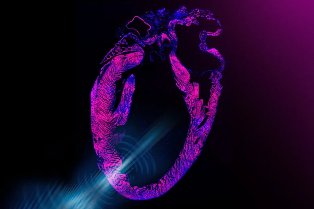
A groundbreaking development from MIT engineers heralds a potential paradigm shift in cardiac care, with the creation of a noninvasive pacemaker that stimulates the heart using precisely directed ultrasound waves. This innovative design could one day offer a much-anticipated surgery-free alternative to the traditional, surgically implanted cardiac devices that millions rely on globally. The research, published in the prestigious journal Nature Biomedical Engineering, outlines a novel approach combining wearable technology with genetic engineering, promising a less intrusive and potentially safer method for managing heart rhythm disorders.
The new device is conceived as a discreet, small sticker designed for adherence to the chest. Embedded within this sticker are miniature transducers that emit focused ultrasound pulses, penetrating the chest wall to interact directly with the heart. At the cellular level, these ultrasound waves are engineered to trigger the opening of specific ion channels within myocardial cells. This critical effect is significantly amplified through a sophisticated genetic engineering technique, ensuring a robust and reliable cellular response. Once these channels open, they facilitate an influx of calcium ions, a fundamental signal that prompts a heart cell to contract and beat, thereby regulating the heart’s rhythm without direct electrical contact.
Addressing the Limitations of Traditional Pacemakers
The development of this ultrasound-based pacemaker addresses long-standing challenges associated with conventional cardiac implants. Currently, approximately 3 million adults in the United States live with pacemakers, and hundreds of thousands more receive new implants each year globally. These small, battery-powered devices are surgically placed in a patient’s chest, with leads extending to the heart to deliver electrical impulses that regulate heart rate. While undeniably life-saving and a cornerstone of modern cardiology, traditional implantable pacemakers are not without their drawbacks.
The surgical implantation procedure, though routine, carries inherent risks such as infection at the implant site, lead dislodgement or fracture, pneumothorax, and hematoma formation. Post-implantation, patients may experience pain, discomfort, or restricted movement. Furthermore, the devices require periodic battery replacement surgeries, typically every 5 to 10 years, which reintroduce surgical risks and patient inconvenience. According to data from the American Heart Association, complications related to lead placement or device infection can occur in 1-5% of patients, sometimes necessitating further surgical interventions or even device extraction. The dream for many years in the medical community has been to achieve effective heart stimulation without these invasive procedures.
Gengxi Lu, a co-corresponding author of the research paper, succinctly articulates this long-held aspiration: "Pacemakers are one of the most important and widely used human implants, and they have saved millions of lives. But they are invasive, and they make direct contact with the beating heart. The dream for many years has been noninvasive heart stimulation with ultrasound." This sentiment underscores the profound impact a truly non-invasive solution could have on patient care and quality of life.
The Science Behind the Breakthrough: Sonogenetics and Ultrasound
The MIT team’s innovation leverages the inherent properties of ultrasound, a form of acoustic wave that can safely penetrate body tissues. For decades, ultrasound has been indispensable in diagnostic imaging, where its waves reflect off internal structures to create detailed images of organs and tissues. More recently, its therapeutic potential has been explored, particularly in neurology, where focused ultrasound is being investigated for treating conditions like Parkinson’s disease, Alzheimer’s, and other brain disorders by modulating neural activity.
Previous studies had already demonstrated that focused ultrasound could activate heart cells in animal models. However, these effects were often inconsistent and relatively weak, lacking the precision and robustness required for clinical pacing. The breakthrough by Zhao and his colleagues lies in their strategic amplification of ultrasound’s effects on the heart, achieved through the burgeoning field of sonogenetics.
Sonogenetics is a relatively nascent approach, drawing conceptual parallels from optogenetics – a revolutionary technique that uses light to control genetically modified cells. In a similar vein, sonogenetics aims to engineer cells to respond specifically and powerfully to sound, including ultrasound. For their ultrasound pacemaker, the team focused on enhancing heart cells’ intrinsic sensitivity to acoustic waves.
In laboratory experiments, the researchers meticulously derived human cardiac cells from embryonic stem cells. These cells then underwent a precise genetic alteration designed to significantly boost their responsiveness to ultrasound. This manipulation specifically led to the production of specialized ion channels that exhibited a much greater propensity to open in the presence of ultrasound waves. As Chen Gong, the paper’s first author, explains, "These channels can now ‘hear’ ultrasound better, and can open to let calcium in, which is what directly activates the cell and causes it to beat." This targeted genetic modification is the linchpin, transforming weak, inconsistent ultrasound responses into a reliable mechanism for cardiac pacing.
From Lab Bench to Prototype: Experimental Validation
The efficacy of this sonogenetically enhanced system was rigorously tested. In experiments involving these engineered human cardiac cells, exposure to ultrasound waves resulted in the cells beating in perfect synchrony with the applied pulses. This synchronized beating was markedly absent in control cells that had not undergone the genetic manipulation, confirming the critical role of sonogenetics.
The team then translated these cellular insights into a tangible device. They fabricated a prototype consisting of an ultrasound sticker, roughly the size of a postage stamp, connected to a small, pocket-sized device containing the necessary batteries and electronics. The sticker itself is made from a proprietary hydrogel material, developed by Zhao’s group, known for its strong adhesion to skin and various materials while simultaneously allowing unimpeded transmission of ultrasound waves. The transducers embedded within the sticker are precisely tunable, enabling them to generate ultrasound waves at specific frequencies tailored for optimal cardiac stimulation.
The next crucial step involved in vivo testing. Researchers administered a sonogenetic, ultrasound-boosting solution, akin to a vaccine, to rats via their tails. This solution introduced the genetic alteration that increased the cardiac cells’ sensitivity to ultrasound. Following this, a miniature version of the pacemaker sticker was adhered to the rats’ chests. Upon activation, the device rapidly and effectively regulated the animals’ heart rates. Rats with bradycardia (slow heart rates) were observed to return to a normal pace, while those experiencing arrhythmias (irregular heartbeats) had their rhythms steadied, beating in perfect sync with the ultrasound "ticks." These results demonstrated the device’s ability to quickly, safely, and noninvasively correct arrhythmias and restore normal, regular heart contractions in a living organism.
A Vision for Integrated Cardiac Care: Monitoring and Stimulation
Beyond mere stimulation, the MIT team envisions a future where this technology offers a comprehensive solution for cardiac management. This same research group previously made headlines for developing adhesive ultrasound stickers capable of imaging deep organs and tissues. Their ambitious next step is to integrate these two functionalities – non-invasive cardiac imaging and non-invasive cardiac stimulation – into a single, unified ultrasound sticker.
Such an integrated device would represent a significant leap forward, enabling simultaneous monitoring and regulation of the heart’s activity in a truly closed-loop system. As Professor Xuanhe Zhao, a professor of mechanical engineering and of civil and environmental engineering at MIT, articulates, "We believe you could one day have stickers on the body that could do long-term imaging deep in the body and also do stimulation for therapeutic effects, in a noninvasive closed-loop way." This closed-loop system would allow the device to continuously assess the heart’s rhythm and immediately intervene with corrective ultrasound pulses if an abnormality is detected, all without patient interaction or invasive procedures.
The Role of Gene Therapy in Future Cardiac Interventions
A key component of the envisioned clinical application for this ultrasound pacemaker is a one-time gene therapy injection. This injection, similar in concept to a vaccine, would deliver the genetic alteration necessary to enhance the sensitivity of cardiac cells to the pacemaker’s ultrasound waves. The concept of gene therapy, while still an evolving field, has gained significant traction and regulatory approval for treating a growing number of inherited conditions, such as sickle cell disease and spinal muscular atrophy, by correcting underlying genetic defects or introducing new functional genes.
The team believes this step would be clinically translatable, providing a crucial enabling technology for noninvasive pacemakers. "We think this step would be clinically translatable as a form of gene therapy that could enable noninvasive pacemakers," Gong emphasizes. This approach bypasses the need for surgical implantation while leveraging established (albeit complex) gene delivery mechanisms. The potential for a single injection to permanently modify cardiac cells to respond to external ultrasound stimulation opens up new avenues for long-term, non-invasive cardiac management.
Collaborative Innovation and Publication
The pioneering research is a testament to collaborative scientific effort. Professor Zhao and his colleagues worked closely with collaborators from Professor Qifa Zhou’s group at the University of Southern California (USC). The multi-institutional team also included researchers from Harvard University, the University of California at Los Angeles, and other groups at USC. The study’s MIT co-authors include first author Chen Gong, along with Runze Li, Won Jun Song, and former postdocs Gengxi Lu, Shucong Li, and Hsiao-Chuan Liu. The publication in Nature Biomedical Engineering underscores the significance and rigor of their findings within the biomedical engineering community.
Broader Implications and the Future of Bioelectronic Medicine
The implications of this non-invasive ultrasound pacemaker extend far beyond simply replacing traditional devices. For millions of patients worldwide suffering from arrhythmias, this technology promises a dramatic improvement in quality of life, eliminating surgical risks, post-operative recovery, and the psychological burden of living with an implanted device. The ability to manage heart conditions with a simple, wearable sticker could democratize access to advanced cardiac care, particularly in regions where surgical facilities are limited.
From an economic perspective, reducing the need for costly surgical procedures, hospital stays, and follow-up interventions could lead to substantial savings for healthcare systems. Furthermore, the concept of a "closed-loop" system, combining continuous monitoring and on-demand stimulation, represents the pinnacle of personalized medicine, potentially preventing severe cardiac events before they escalate.
This breakthrough also energizes the broader field of bioelectronic medicine, which seeks to use technology to modulate biological processes. The successful application of sonogenetics in cardiac pacing could inspire similar approaches for other organs and conditions. As Professor Zhao states, "In this paper, we demonstrated noninvasive pacemaking. However, we think this concept could be useful beyond just the heart. We believe you could one day have stickers over different parts of the body that could do long-term imaging, monitoring, and closed-loop therapeutic stimulation." This vision encompasses applications ranging from nerve stimulation to organ function regulation, opening up a vast landscape for future medical innovation.
While clinical translation will undoubtedly involve extensive further research, including larger animal studies and human trials, the foundational work by the MIT team has laid a robust groundwork. Challenges will include optimizing gene therapy delivery for human safety and efficacy, ensuring the long-term stability and biocompatibility of the sticker, and navigating complex regulatory pathways. However, the promise of a surgery-free future for cardiac care, powered by the synergy of ultrasound and genetic engineering, offers profound hope and a compelling direction for the next generation of medical technology.
This groundbreaking work received crucial support from several prominent institutions, including the National Institutes of Health, the National Science Foundation, the Department of Ophthalmology from Research to Prevent Blindness, and the U.S. Department of War, highlighting the interdisciplinary and strategic importance of this research.