In a landmark achievement for the field of ultrafast photochemistry, an international team of researchers has successfully observed the intricate movement of energy through a molecule in the immediate aftermath of light absorption. Utilizing the world-class capabilities of the European X-ray Free-Electron Laser (European XFEL), the scientists were able to demonstrate that individual atoms within a single molecule act as distinct reporters, recording vastly different aspects of the molecular transformation. This breakthrough, achieved through the use of rapid X-ray flashes, offers a high-definition view of how energy is redistributed across atomic structures on timescales measured in trillionths of a second.
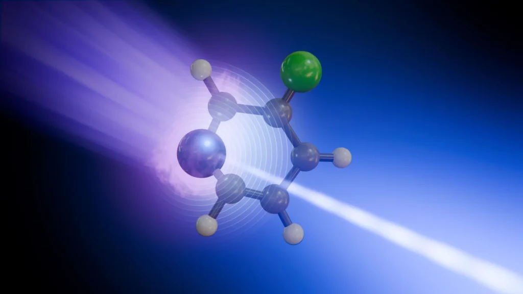
The study, which focused on the molecule 3-fluoropyridine, reveals that exposure to light does not merely excite a molecule as a whole but can make specific atoms more responsive to the movements of their neighbors. This "site-specific" sensitivity allows researchers to decouple electronic changes from structural vibrations, providing a roadmap for understanding how complex chemical systems handle energy. The findings have profound implications for a variety of scientific disciplines, ranging from the development of more efficient solar energy harvesting materials to understanding the fundamental mechanisms that protect biological molecules like DNA from radiation damage.
The Mechanism of Ultrafast Photochemistry
When a molecule absorbs a photon, particularly in the ultraviolet (UV) spectrum, it undergoes a rapid transition from its stable "ground state" to an "excited state." This process is the starting point for nearly all light-driven chemistry, including photosynthesis in plants and the visual cycle in human eyes. However, the excited state is inherently unstable. To return to stability, the molecule must dissipate this excess energy, often through a combination of structural deformation and electronic relaxation.
The researchers selected 3-fluoropyridine for their study because of its specific geometric and electronic properties. A small, ring-shaped molecule consisting of carbon, hydrogen, nitrogen, and a single fluorine atom, 3-fluoropyridine serves as an ideal model for more complex heterocyclic compounds found throughout nature. Under normal conditions, the molecule maintains a flat, planar structure. However, the team discovered that upon absorbing a brief pulse of ultraviolet light, the molecule’s electrons jump to a higher energy level, triggering an immediate and violent bending of the molecular frame.
As the molecule distorts, it encounters a "conical intersection." In the world of quantum chemistry, conical intersections are treated as high-speed "funnels" where the energy levels of different electronic states meet. At these points, the traditional separation between electronic motion and the movement of the atomic nuclei—known as the Born-Oppenheimer approximation—breaks down. This coupling allows for the lightning-fast transfer of energy from the electrons to the structural skeleton of the molecule, effectively converting potential electronic energy into kinetic vibrational energy.
A Chronology of Molecular Transformation
The experimental process conducted at the European XFEL followed a precise chronological sequence, often referred to as a "pump-probe" technique. This method allows scientists to take "snapshots" of a reaction at different stages of its evolution.
- The Pump Phase: The process began with an ultrashort ultraviolet laser pulse. This pulse acted as the "pump," delivering a specific amount of energy to the 3-fluoropyridine molecules and initiating the excitation process. This set the "time zero" for the experiment.
- The Interaction Phase: Within femtoseconds (quadrillionths of a second) of the UV pulse, the molecule began to exit its planar shape. The electronic distribution shifted, and the molecule approached the conical intersection.
- The Probe Phase: To observe what was happening at specific moments after the excitation, the researchers used a second pulse: a soft X-ray flash produced by the European XFEL. This "probe" pulse was timed to arrive at delays ranging from a few femtoseconds to several picoseconds after the initial UV pump.
- Ionization and Detection: The X-ray pulse was energetic enough to remove deeply bound "core" electrons from either the nitrogen or the fluorine atoms within the molecule. By measuring the kinetic energy of these ejected electrons—a technique known as time-resolved X-ray photoelectron spectroscopy (tr-XPS)—the researchers could determine the exact chemical and electronic environment of that specific atom at that exact moment.
- Reconstruction: By repeating this measurement thousands of times with different delay intervals, the team reconstructed a frame-by-frame "movie" of the energy flow through the molecule.
Site-Specific Reporting: Nitrogen vs. Fluorine
One of the most significant findings of the study was the divergence in the "stories" told by the different atoms. Despite being part of the same small molecule, the nitrogen and fluorine atoms provided entirely different perspectives on the energy dissipation process.
The nitrogen atom, which is integrated directly into the molecular ring, was found to be a highly sensitive reporter of the initial electronic excitation. Because the nitrogen atom’s lone pair of electrons is heavily involved in the molecule’s transition to an excited state, the signals captured from the nitrogen site reflected a complex mixture of electronic redistribution and the initial structural bending.
In contrast, the fluorine atom, which sits on the periphery of the ring, acted as a cleaner indicator of the molecule’s vibrational relaxation. The fluorine signal was less affected by the initial electronic "chaos" and instead provided a clear timeline of how the vibrations, triggered by the crossing of the conical intersection, eventually settled down or "relaxed" across the molecular structure.
Antonio Picón, a co-author of the study from the Instituto de Ciencia de Materiales de Madrid (ICMM-CSIC), emphasized the importance of this distinction. "We can now see that not every atomic site tells the same story in the signals we capture from our X-ray pulses," Picón noted. "Some atoms report where the charge is going, while others reveal how the whole molecule vibrates."
Technical Prowess of the European XFEL
The success of this experiment was predicated on the unique capabilities of the European XFEL, located in the Hamburg area of Germany. The facility features a 3.4-kilometer-long tunnel housing a superconducting linear accelerator that accelerates electrons to near-light speeds. These electrons are then directed through undulators—series of magnets with alternating poles—which force the electrons onto a zigzag path, causing them to emit extremely intense, short-lived X-ray pulses.
The specific instrument used in this study was the Small Quantum Systems (SQS) instrument. SQS is designed specifically to study the fundamental processes of light-matter interaction in atoms, molecules, and clusters. The high brightness of the X-ray pulses allowed the team to capture signals from dilute gas-phase molecules with unprecedented clarity.
Furthermore, the research required advanced computer simulations to interpret the raw data. Because the quantum mechanics of a molecule passing through a conical intersection are incredibly complex, the team utilized theoretical models to connect the observed electron energies with the underlying physical movements. These simulations confirmed that the "chemical shifts" seen in the X-ray photoelectron spectra were indeed the result of the molecule’s rapid change in shape and electronic state.
Scientific Context and Broader Implications
The ability to watch chemical changes at specific atomic sites on their natural timescale—the femtosecond and picosecond range—is a long-held dream of chemists. For decades, most spectroscopic techniques provided an "averaged" view of a molecule, making it difficult to determine which specific part of a structure was responsible for a particular behavior.
The findings from this study have immediate relevance for several key areas of research:
1. Photoprotection in Biological Systems: One of the great mysteries of biology is how DNA remains stable despite constant exposure to UV radiation from the sun. It is known that DNA bases possess "ultrafast relaxation" mechanisms that convert potentially damaging UV energy into harmless heat before chemical bonds can break. The techniques used at the European XFEL could eventually allow scientists to map these pathways in DNA, revealing the exact "safety valves" that prevent genetic mutations.
2. Artificial Photosynthesis and Solar Energy: In the quest for better renewable energy, scientists are designing synthetic molecules that can capture sunlight and transport that energy to a reaction center. Understanding the "bottlenecks" where energy is lost or diverted is crucial for increasing the efficiency of these materials. By observing energy flow at the atomic level, engineers can design molecules that guide energy more effectively toward its destination.
3. Next-Generation Materials: Many modern technologies, from organic LEDs (OLEDs) to high-speed optical switches, rely on the interaction of light and matter. This research provides a new toolkit for materials scientists to observe how these interactions occur in real-time, potentially leading to the development of faster and more energy-efficient electronic components.
Official Responses and the Future of the Field
The international collaboration included researchers from various prestigious institutions, highlighting the global importance of the work. Daniel Rivas, a former instrument scientist at SQS and now a guest scientist at the facility, highlighted the strategic value of the experiment.
"This is what European XFEL was built to enable: watching chemical change where it begins, at specific atomic sites and on its natural timescale," Rivas said. "By combining multi-site sensitivity with femtosecond resolution, we are opening a new window on the microscopic mechanisms that govern photochemistry."
The research team plans to extend this approach to increasingly complex systems. While 3-fluoropyridine is a relatively simple molecule, the "site-specific" methodology is scalable. Future experiments may target larger biomolecular building blocks or functional organic molecules used in medicine.
The study also underscores the growing synergy between experimental physics and theoretical chemistry. As X-ray sources become more powerful and pulses become even shorter (reaching the attosecond range), the data generated will require even more sophisticated computational tools to decode. This "virtuous cycle" of better experiments and better theory is expected to accelerate the pace of discovery in the molecular sciences over the coming decade.
In conclusion, the observation of energy redistribution in 3-fluoropyridine marks a significant milestone in our ability to probe the quantum world. By turning individual atoms into microscopic witnesses, scientists have gained a new level of control and understanding over the fundamental processes that drive chemistry, biology, and materials science. The "new window" opened by the European XFEL promises to reveal a wealth of hidden mechanisms that have, until now, occurred too fast and on too small a scale for the human eye—or even the most advanced conventional microscopes—to see.