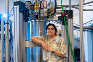
A groundbreaking discovery by researchers at the Massachusetts Institute of Technology (MIT), led by Associate Professor Ritu Raman, has unveiled a novel method for engineering and precisely controlling the growth patterns of new blood vessels. Published in the prestigious Proceedings of the National Academy of Sciences, their findings demonstrate that simple mechanical stimulation—repeatedly jostling or stretching an artery—can induce the sprouting of smaller capillaries and dictate their direction and density. This innovative approach promises to overcome significant hurdles in the field of regenerative medicine, offering a scalable and reproducible pathway to fabricating organized blood vessel networks within engineered tissues, a critical step toward creating functional artificial organs and repairing damaged tissues in the human body.
The Critical Challenge of Vascularization in Tissue Engineering
The ability to grow new, functional tissues and organs outside the body for transplantation or disease modeling has long been a holy grail in biomedical science. However, a persistent and formidable challenge in tissue engineering has been the lack of effective strategies to create robust, intricate vascular networks that can supply engineered tissues with oxygen and nutrients, and remove waste products. Without such a network, cells within larger engineered constructs quickly starve and die, limiting the size and complexity of tissues that can be successfully developed.
Millions worldwide suffer from conditions that could benefit from advanced tissue engineering. Heart disease remains the leading cause of death globally, often involving damage to cardiac tissue that current therapies struggle to fully repair. Diabetes frequently leads to complications like chronic non-healing wounds and organ damage, where enhanced vascularization could prove life-saving. Organ failure, from liver to kidney, necessitates transplantation, but the scarcity of donor organs underscores the urgent need for lab-grown alternatives. Trauma, burns, and neurodegenerative diseases also present scenarios where the ability to regenerate functional, vascularized tissue could dramatically improve patient outcomes.
Current approaches to vascularizing engineered tissues have faced significant limitations. While 3D printing technologies have advanced to create larger structures, they struggle to replicate the delicate, microscopic networks of capillaries essential for tissue viability. These capillaries, often just a few micrometers in diameter, are too fine and intricate for current additive manufacturing techniques. Another strategy involves using chemical cues, such as growth factors, to stimulate vessel growth. While effective to some extent, these factors are difficult to pattern precisely, degrade quickly, can be expensive, and may induce unwanted off-target effects. Controlling the spatial organization and density of new vessels using chemical gradients alone has proven largely impractical for complex tissue architectures. As Professor Raman notes, "You can try to pattern chemical cues, like growth factors, to direct where vessels grow, but you can’t do this very precisely. We thus need other types of patternable cues that can help us build tissues with organized vessels."
A Paradigm Shift: Mechanical Forces as Biological Architects
The MIT team’s breakthrough pivots from purely chemical or structural approaches to leverage the inherent mechanosensitivity of biological systems. Their work builds upon a foundational understanding that mechanical forces play a crucial role in regulating cellular behavior, tissue development, and disease progression within the body. Blood vessels, in particular, are constantly subjected to mechanical stresses from blood flow (shear stress) and surrounding tissue (tensile and compressive forces), which influence their growth, remodeling, and maintenance.
This new research is a direct continuation of Raman’s lab’s prior success in using mechanical stimulation to guide the growth of artificial muscles and nerves. In earlier experiments, the team developed a "wobbly gel mat" system where muscle or neuron cells were grown on a nutrient-infused gel containing a small embedded magnet. By manipulating an external magnet, they could mechanically "exercise" the cells, pulling the cell-covered gel back and forth. This demonstrated that physical exertion directly influenced how these cells grew and organized, leading to more functional muscle fibers and improved neuronal connections. The success of these previous endeavors laid the groundwork for investigating whether similar mechanical principles could be applied to the notoriously challenging task of vascularization.
The "Blood-Vessel-on-a-Chip" Innovation and Precision Control
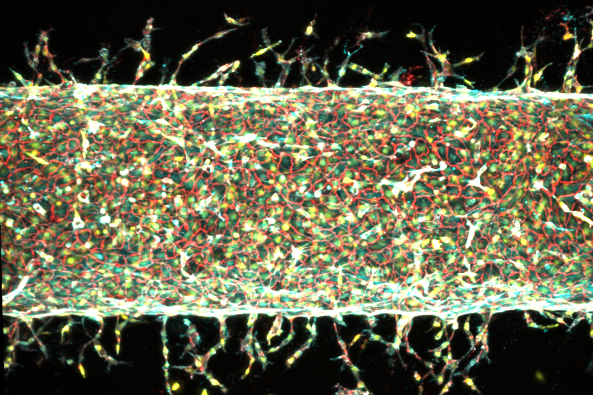
To test their hypothesis, Raman and her team engineered a sophisticated "blood-vessel-on-a-chip" device. This microfluidic platform, smaller than a postage stamp, was filled with a specialized nutrient-rich gel designed to mimic the extracellular matrix found in the body. A thin channel was created through the gel, which was then coated with live endothelial cells. These cells, which naturally line the interior of blood vessels, spontaneously organized to form a central, artery-like structure within the channel, and began to sprout new, capillary-like vessels into the surrounding gel.
The ingenious aspect of the setup involved integrating magnetic manipulation. A small magnet was embedded within the gel, and the entire device was placed under a motorized stage fitted with external magnets. By precisely moving these external magnets, the researchers could apply controlled mechanical forces—jostling and stretching—to the central artery and its nascent capillary network. This allowed them to observe, in real-time and with remarkable precision, how the blood vessels responded to varying degrees and directions of mechanical stimulation.
The results were compelling and demonstrated an unprecedented level of control over vascular growth:
- Enhanced Sprouting: When the central artery was repeatedly jostled, a significantly greater number of new capillaries sprouted from its length compared to control arteries left undisturbed. This confirmed the initial hypothesis that "moving is good" for vascular growth.
- Directional Guidance: By changing the direction in which the gel was stretched, the researchers could precisely redirect the growth of the new vessels. The capillaries would follow the imposed mechanical pattern, turning and growing along the lines of mechanical tension. This represents a critical step towards engineering complex, organized vascular networks rather than random growth.
- Quantitative Control of Sprouting: The degree of stretching also influenced the characteristics of the new vessels. A moderate stretch, around 5 percent of the gel’s total width, promoted the growth of many new vessels. Conversely, a more significant stretch, around 15 percent, resulted in fewer but longer vessels. This fine-tuning capability provides engineers with a powerful tool to program specific vascular architectures tailored to different tissue types and functional requirements.
"We’re finding that moving is good, which is always the takeaway of everything we do in our lab," Raman stated, underscoring the consistent theme of mechanical forces driving biological processes in her research. "Mechanical forces play an important role in our bodies. That means that if you want to grow more or less vessels, or shorter or longer vessels, or vessels in certain directions, we now know how to do that."
Unveiling the Molecular Mechanism: The Role of Piezo1
Beyond demonstrating control, the team delved deeper to understand the underlying biological mechanisms. This pursuit led them to investigate the role of a specific gene: Piezo1. The inspiration for this line of inquiry came from a talk by molecular biologist Ardem Patapoutian, who was awarded the Nobel Prize in Physiology or Medicine in 2021 for his seminal discovery of mechanosensitive ion channels.
Patapoutian’s work identified PIEZO1 and PIEZO2 as critical ion channels embedded in cell membranes that act as gatekeepers, opening and closing in response to mechanical pressure. These channels convert physical stimuli into electrochemical signals, influencing a myriad of cellular processes. After Raman presented her team’s preliminary findings on mechanical stimulation and blood vessel growth to Patapoutian, he proposed that the PIEZO1 channel might be the key. The hypothesis was that the mechanical exercise of the central artery could be activating these ion channels in the endothelial cells, thereby triggering the cascade of events leading to new blood vessel formation.
To rigorously test this hypothesis, Raman’s team employed gene editing techniques to suppress the PIEZO1 gene in the endothelial cells. They then repeated their mechanical stimulation experiments. The results were striking: with the PIEZO1 gene less active, significantly fewer new blood vessels sprouted, even when the central artery was subjected to the same mechanical exercise. This crucial finding confirmed that PIEZO1 channels are indeed essential mediators in the mechanotransduction pathway that translates physical force into vascular growth. This discovery not only explains how mechanical forces drive angiogenesis but also identifies a potential molecular target for future therapeutic interventions.
Broader Impact and Transformative Implications for Medicine
The implications of this research extend across multiple frontiers of biomedical science and regenerative medicine. The ability to program blood vessel growth with physical cues represents a paradigm shift with the potential to revolutionize how we approach tissue engineering, disease modeling, and clinical therapies.
-
Regenerative Medicine and Organ Transplantation: The most immediate and profound impact is on the creation of functional, transplantable tissues and organs. Currently, the lack of vascularization severely limits the thickness and viability of engineered tissues. This new method could enable the fabrication of pre-vascularized grafts for a range of organs, including heart muscle, liver tissue, kidney components, and even complex skin grafts for burn victims. Such tissues would be ready to integrate into the body and establish immediate blood flow, significantly improving their chances of survival and function post-implantation. For instance, imagine generating a patch of heart muscle with its own integrated blood supply, ready to be sutured onto a damaged heart.
-
Disease Modeling and Drug Discovery: The "blood-vessel-on-a-chip" platform itself, enhanced with programmable vascularization, offers a superior model for studying diseases. Researchers could create more realistic "organs-on-a-chip" that accurately mimic human physiology, including blood flow dynamics. This would allow for more effective screening of new drugs, understanding disease progression (e.g., atherosclerosis, tumor angiogenesis), and investigating the effects of various therapies in a controlled, in-vitro environment, reducing reliance on animal testing.
-
Wound Healing and Trauma Repair: Chronic wounds, particularly those associated with diabetes or poor circulation, often fail to heal due to inadequate blood supply. The ability to stimulate and direct new capillary growth could lead to novel therapies for accelerating wound closure and improving tissue regeneration in traumatic injuries or reconstructive surgeries.
-
Cancer Research: Angiogenesis, the formation of new blood vessels, is a critical process in tumor growth and metastasis. Understanding and controlling vessel growth using mechanical cues could offer new avenues for anti-angiogenic therapies that starve tumors of their blood supply, or conversely, for revascularizing tissues damaged by radiation or chemotherapy.
-
Personalized Medicine: In the long term, this technology could contribute to personalized medicine, where patient-specific cells are used to engineer tissues that are genetically matched, minimizing immune rejection and maximizing therapeutic efficacy.
Challenges and Future Directions
While immensely promising, this research represents a foundational step. Several challenges and future directions lie ahead. Scaling up the process from postage-stamp-sized chips to larger, clinically relevant tissue constructs will require significant engineering innovation. Integrating these precisely vascularized networks with other cell types (e.g., muscle cells, nerve cells) to create multi-tissue organoids is another complex task. Long-term stability and functionality of the engineered vessels will need thorough investigation. Furthermore, rigorous in-vivo testing in animal models will be necessary to confirm the safety and efficacy of these mechanically guided vascular networks before any human applications can be considered.
As co-author Jessica Shah states, "We are now investigating how precisely patterning blood vessel growth can help improve muscle function," highlighting the immediate next steps for the team. This iterative process of discovery, mechanistic understanding, and application is characteristic of translational research.
The work was supported, in part, by critical funding from the U.S. Department of War Army Research Office Early Career Program and PECASE Grant, and a Department of War DURIP Program Grant, underscoring the potential military applications in treating battlefield injuries and advancing trauma care.
This breakthrough from MIT marks a pivotal moment in tissue engineering. By harnessing the power of mechanical forces and elucidating the role of key molecular players like Piezo1, researchers have unlocked a new dimension of control over one of biology’s most fundamental and elusive processes: the precise formation of blood vessel networks. This innovation not only deepens our understanding of mechanobiology but also provides a concrete, programmable pathway toward a future where engineered tissues and organs can reliably restore function and improve human health.