Epithelial cells, the ubiquitous guardians forming the protective linings and coverings throughout our bodies, have long been recognized for their critical roles in safeguarding our internal and external environments. From orchestrating the intricate dance of embryonic development to diligently repairing the most minor skin scrape or major surgical incision, these cells are fundamental to life itself. They constitute the outer layer of our skin, the delicate surfaces of our organs, and the crucial inner lining of blood vessels, acting as both structural components and dynamic responders to physiological demands. However, when the finely tuned regulatory mechanisms governing epithelial cell behavior falter, these essential cells can undergo malignant transformation, leading to uncontrolled proliferation and the ominous spread of cancer throughout the body.
A groundbreaking study by engineers at the Massachusetts Institute of Technology (MIT) has now revealed a previously unobserved, collective behavior among migrating epithelial cells: a synchronized, rhythmic pulsing. Published today in the esteemed journal Newton, this research describes groups of epithelial cells repeatedly converging and then receding, akin to a circle of dancers elegantly moving together and then pulling apart. This remarkable discovery adds a new dimension to our understanding of cellular dynamics, shifting focus from purely spatial coordination to the temporal orchestration of cell movements.
The research team, led by Professor Ming Guo of mechanical engineering at MIT, meticulously measured this collective rhythmic pulsing across various types of epithelial cells. Their investigations included healthy cells, cells derived from benign tumors, and those from aggressive cancerous growths. The findings presented a striking and potentially clinically significant pattern: malignant epithelial cells exhibited a notably more persistent synchronization, maintaining their collective pulsing for approximately twice as long as their healthier counterparts. While the precise biological mechanisms driving this synchronized "cellular dance" remain to be fully elucidated, the researchers propose that this rhythmic coordination could serve as a vital clinical signal, offering new avenues for early detection and therapeutic intervention.
Professor Guo emphasized the potential implications of this discovery, stating, "More aggressive cancer cells tend to have a steadier and more persistent rhythm as compared to healthy ones. We think this coordination could serve as an early warning sign of how likely a tumor is to spread." He further hypothesized that the same coordinated waves observed in the lab might play pivotal roles in the natural shaping of embryos during development and the efficient closure of wounds following injury, highlighting the fundamental nature of this newly identified cellular behavior across diverse biological processes. The study’s authorship includes first author Wenhui Tang, a former MIT graduate student, alongside Mehrana Nejad and L. Mahadevan of Harvard University, and Adrian Pegoraro of the Metrology Research Centre of the National Research Council Canada, underscoring the collaborative and interdisciplinary nature of this pioneering work.
The Unseen Symphony: Deciphering Temporal Cell Coordination
For decades, scientific inquiry into how epithelial cells organize themselves to form complex organs and tissues has largely concentrated on spatial coordination. Researchers have meticulously mapped where cells move, their relative positions within growing tissues, and their ultimate destinations. These spatial relationships are undeniably critical for understanding morphogenesis and tissue homeostasis. However, the temporal dimension—how cellular movements and interactions evolve over time in a coordinated fashion—has remained less explored, representing a significant frontier in cell biology.
Professor Guo’s group at MIT specializes in investigating cell interactions to identify distinctive patterns indicative of healthy versus diseased states. Their approach often involves capturing microscopic snapshots of cells cultured in the laboratory, allowing them to observe and analyze intriguing cellular behaviors. It was within this context that Wenhui Tang, then a member of Guo’s lab, made her pivotal observation. While reviewing a series of time-lapse movies of epithelial cells, Tang began to discern a subtle yet distinct rhythm, a repetitive pattern unfolding over time.
"I was studying collective cell migration, and I observed cells were swelling, then squeezing together, then swelling, again and again, forming local patterns," Tang recalled. "That’s when I realized there might be something more interesting happening with these cells over time." This serendipitous observation laid the foundation for the rigorous investigation into the temporal coordination of epithelial cells, challenging existing paradigms and opening new avenues for research into fundamental biological processes and disease mechanisms.
Methodology: Capturing the Cellular Pulse
To systematically investigate this rhythmic cellular movement, the researchers designed a meticulous experimental protocol focusing on the precise timing of cellular movements. They began by culturing healthy, live epithelial cells, specifically MDCK (Madin-Darby Canine Kidney) cells, a widely used model in cell biology for studying epithelial monolayers and cell migration. To make individual cells distinguishable and track their movements, they stained the cell nuclei with a fluorescent dye. This allowed for clear identification and tracking of each cell within the larger collective. The cells were maintained in nutrient-rich dishes, providing an optimal environment for their natural growth, division, and migration.
Professor Guo elaborated on the experimental design: "We’re looking at their natural migration process, related to how they would migrate during different processes in the body, such as when forming skin and organs, and healing wounds." This emphasis on natural migration underscores the physiological relevance of their observations. Using a sophisticated confocal microscope, the team captured high-resolution images of the cells every few minutes, extending the observation period for up to 30 hours. The subsequent compilation of these sequential images into time-lapse movies revealed a distinct and undeniable pattern.
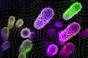
"If you just stare at any one location, you can see those dots are coming together, and then going further away, then coming together again, and going further away, like waves," Tang described, vividly illustrating the observed phenomenon. The analysis showed that a single pulse, characterized by cells moving inward and then outward, transpired over approximately one hour. This pulsing behavior was observed to persist in healthy cells, maintaining a slow and steady rhythm throughout the entire 30-hour observation period. The rhythmic nature of this collective movement suggested an underlying regulatory mechanism, prompting further investigation into its implications for disease.
Malignancy’s Persistent Rhythm: A Potential Biomarker
Intrigued by the consistent pulsing in healthy cells, the team extended their investigation to ascertain whether other types of epithelial cells, particularly those associated with disease, would exhibit similar synchronization. They conducted the same experiment using several different lines of human breast cancer epithelial cells, encompassing cells from benign tumors and those displaying increasing degrees of malignancy. The results were compelling: similar pockets of synchronized pulsing were observed in every cell type studied, but critically, this phenomenon was most pronounced and persistent in the most cancerous cells.

"We found the really dangerous cancer cells team up over time, and do this persistent oscillation, twice as long as healthy cells," Professor Guo revealed. "This is unexpected. We see they really team up, synchronize, and oscillate together, which potentially facilitates their invasion." This observation is profoundly significant because collective cell migration is a well-established mechanism through which cancer cells disseminate from primary tumors to establish metastatic lesions, which are responsible for the vast majority of cancer-related deaths. The enhanced, persistent synchronization in malignant cells suggests a refined, coordinated strategy that could confer an advantage in their invasive journey.
The researchers also uncovered a fascinating correlation between cell synchronization and cell density. In each dish of cells, regardless of their type, the cells continuously grew, divided, and pulsed. As their numbers increased, more cells began to pulse together, leading to a measurable increase in their collective synchronization. However, this trend was not indefinite. Once the cells reached a certain critical density, their pulsing activity began to diminish. "There’s a peak of synchrony before it decreases as cell density continues to increase," Tang explained. This intricate relationship between density and synchronization hints at complex feedback mechanisms that govern collective cellular behavior, possibly involving mechanosensing and contact inhibition.
Broader Implications: From Asthma to Cancer Therapy
The implications of this discovery extend far beyond merely understanding cell movement, offering potential breakthroughs in diverse areas of medicine, from chronic respiratory conditions to the daunting challenge of cancer.
1. Early Cancer Detection and Prognosis:
The finding that more aggressive cancer cells exhibit a more persistent and steady rhythm suggests that this collective pulsing could serve as a novel biomarker for cancer progression and metastatic potential. Current diagnostic methods for cancer often rely on morphological changes, genetic mutations, and protein markers. A dynamic, biophysical marker like synchronized cellular pulsing could provide complementary, real-time insights into tumor aggressiveness. For instance, analyzing biopsy samples or even circulating tumor cells for this specific rhythmic signature could potentially help clinicians identify patients at higher risk of metastasis, allowing for more aggressive or tailored treatment strategies earlier in the disease course. While further research is needed to validate its clinical utility, the concept offers a tantalizing new avenue for prognostic assessment.
2. Novel Therapeutic Targets for Cancer:
If the underlying molecular and biophysical mechanisms responsible for this synchronized pulsing in malignant cells can be precisely identified, they could represent new therapeutic targets. Imagine drugs designed to specifically disrupt this coordinated cellular rhythm, thereby hindering the collective migratory capabilities of cancer cells and potentially inhibiting their ability to invade surrounding tissues and metastasize. Professor Guo envisions such a future: "More malignant cells would be better synchronized. After treating them with a drug, if their synchronization is disrupted, then it might be an efficient drug where we can consider the next step." This offers a functional screening approach for anti-cancer agents, moving beyond simply killing cells to disrupting their coordinated destructive behaviors.
3. Insights into Developmental Biology:
The hypothesis that these coordinated waves may help shape embryos during development underscores the fundamental nature of this cellular rhythm. Embryogenesis is a marvel of precise cellular orchestration, where cells migrate, differentiate, and organize into complex structures with exquisite timing. Understanding how collective pulsing contributes to processes like gastrulation, neural tube closure, or organogenesis could shed new light on developmental disorders and provide a deeper understanding of the physical forces that sculpt life forms.
4. Advancements in Wound Healing:
Similarly, the role of synchronized pulsing in closing wounds upon injury suggests that manipulating this rhythm could enhance wound repair. Chronic wounds, which affect millions globally, are a significant clinical challenge. If epithelial cells use this coordinated movement to efficiently bridge gaps and restore tissue integrity, then fostering or enhancing this synchronization through topical treatments or cellular therapies could accelerate healing, reduce scarring, and prevent complications. This perspective opens up avenues for regenerative medicine and tissue engineering.
5. Targeting Epithelial-related Diseases like Asthma:
The observed connection between cell density and synchronization holds particular relevance for conditions affecting epithelial barriers, such as asthma. Epithelial cells form the crucial lining of airways. In healthy individuals, these cells pack tightly together, forming a robust, "jammed" barrier that protects the airways from irritants and pathogens. In asthmatic airways, however, this epithelial layer is often compromised, becoming looser and more fragile, making it easily irritated and difficult to heal. The research suggests that altered cell density and synchronization could contribute to this compromised barrier function.
By understanding how potential drugs affect asthma cell synchronization, researchers might be able to identify novel treatments that restore the proper "jamming" state of airway epithelial cells. This could lead to therapies that not only alleviate symptoms but also address the underlying epithelial dysfunction in asthma, offering a more targeted and effective approach to disease management. Similar implications could extend to other diseases characterized by compromised epithelial barriers, such as inflammatory bowel disease or certain dermatological conditions.
The Road Ahead: From Observation to Intervention
The MIT study represents a pivotal moment in cellular biology, transitioning from primarily spatial analyses of cell migration to an appreciation of its temporal dynamics. The discovery of synchronized pulsing in epithelial cells, particularly its enhanced persistence in malignant cells, opens up a wealth of research opportunities. Future investigations will undoubtedly focus on elucidating the precise molecular machinery and biophysical forces that drive this rhythmic behavior. Understanding the signaling pathways, cytoskeletal components, and cell-cell adhesion molecules involved will be crucial for translating these fundamental insights into practical clinical applications.
This interdisciplinary research, supported in part by the National Institutes of Health, highlights the power of combining engineering principles with biological inquiry. By applying quantitative analysis and sophisticated microscopy techniques, the MIT team has unveiled a hidden layer of cellular communication and coordination. As scientists continue to unravel the complexities of this cellular dance, the promise of new diagnostic tools, targeted therapies, and a deeper understanding of life’s fundamental processes moves ever closer to realization. The rhythm of life, it appears, holds secrets yet to be fully uncovered, offering hope for a future where diseases are understood and treated with unprecedented precision.