While the sheer power required for human swimming is evident in every stroke of an Olympic athlete, a groundbreaking discovery by engineers at the Massachusetts Institute of Technology (MIT) reveals that even a single, meticulously designed layer of muscle cells can generate enough force to propel a robot through water. This significant advancement, detailed in a paper published today in the prestigious journal Advanced Functional Materials, introduces a novel design for a thin, muscle-powered swimming robot, marking a critical step forward in the nascent field of biohybrid robotics.
The core of this innovative aquabot lies in its unique construction: a minimalist "skeleton" crafted from a film of gel, approximately the length and width of a standard stick of chewing gum. This gel film is ingeniously divided into two halves, each forming a "fin" for the robot. Crucially, each fin is adorned with a layer of live muscle cells, astonishingly thin—far less than the diameter of a single human hair. These specialized muscle cells are not just any cells; they have been genetically engineered to exhibit a remarkable property: they twitch and contract in direct response to pulses of light, a technique known as optogenetics.
The operational mechanism is elegantly simple yet profoundly effective. When researchers direct a beam of light onto one of the robot’s fins, the genetically modified muscle cells on its surface instantly react, contracting in a synchronized twitch. This contraction causes the entire fin to flap with sufficient force to push the robot through its aquatic environment. By precisely controlling the light exposure—flashing it on one fin or the other, and varying the intervals and intensity—the engineers can meticulously control the swimming robot’s direction and speed, enabling sophisticated maneuverability.
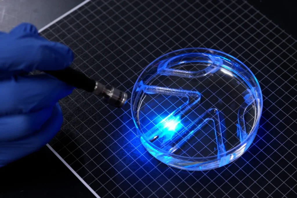
In initial demonstrations, this paper-thin marvel proved its capabilities by successfully navigating and swiveling through a basic watery maze within a petri dish. Performance metrics show the robot achieving a maximum speed of approximately four times its own body length per minute. While this pace might seem modest when compared to the blistering speed of Olympic swimmers, who can cover up to 65 body lengths per minute, it is remarkably competitive with the more leisurely aquatic explorers of the natural world, such as the cow shark, which cruises through the ocean at a comparable rate. "It takes a lot of force to move through water versus air," notes Ritu Raman, an associate professor of mechanical engineering at MIT and a lead author of the study, emphasizing the impressive strength of the robot relative to its diminutive size. "The robot’s quite strong, given its size."
A New Dimension in Biohybrid Robotics: The 2D Breakthrough
This new robot stands as a pivotal achievement, representing the first instance of a very thin, two-dimensional, muscle-powered robot capable of sustained locomotion. Its design philosophy starkly contrasts with many preceding biohybrid robot prototypes, which typically rely on bulky, three-dimensional chunks of lab-grown skeletal muscle. Such conventional designs often necessitate millions of cells for their fabrication, making them resource-intensive and often less agile.
Professor Raman highlights the distinct advantages of this thinner, less bulky design. "Currently, biohybrid robots from our group and others are built from bulky, 3D chunks of lab-grown skeletal muscle that require millions of cells to fabricate," she explains. The team’s novel, paper-thin bot promises significant benefits, including lower construction costs and enhanced movement efficiency. Beyond these practical considerations, Raman envisions a future where biohybrid robots, powered by living muscle, could undertake delicate tasks in environments too fragile or unpredictable for conventional, rigid hardware. The inherent properties of living tissue—its softness, responsiveness to surroundings, and remarkable capacity for self-healing—make it an ideal candidate for such sensitive applications, opening doors to previously unimaginable possibilities in exploration and intervention.
The collaborative effort behind this research involved a dedicated team of MIT co-authors, including first author Maheera Bawa, Arielle Berman, Laura Schwendeman, Ferdows Afghah, and Seanbiron Johnson, underscoring the interdisciplinary nature of this scientific endeavor.
The Evolution of Biohybrid Actuators: A Chronological Journey
The development of this advanced swimming robot is not an isolated breakthrough but rather the culmination of years of focused research within Professor Raman’s group, building upon prior innovations in bio-inspired actuation. The journey to maximize muscle movement for robotics began with fundamental investigations into how living cells could be harnessed to generate controllable mechanical force.
A significant precursor to the current swimming robot was the group’s work last year, which resulted in an iris-inspired disk of artificial muscle tissue. In that project, researchers ingeniously stamped a disk of gel with a pattern of concentric and radial grooves. Live muscle cells were then carefully deposited onto the gel’s surface, where they naturally aligned and grew along these predetermined grooves. When stimulated with light, these cells moved in complex patterns, causing the disk to stretch and squeeze, mimicking the intricate dilation and constriction of a human iris in response to light.
That earlier work was groundbreaking in demonstrating that muscle cells could be grown in remarkably thin layers and arranged in complex patterns, which, when stimulated, could produce controllable, multi-directional movements. "People hadn’t seen this muscle architecture engineered from scratch before," Raman recounted, highlighting the novelty of their cellular arrangement. "And the cells were moving in multiple directions." However, despite the innovative architecture, the movements generated were relatively small, typically around 100 microns. "From a robotics perspective, their movements were tiny," Raman acknowledged, identifying the primary limitation that needed to be overcome for practical robotic locomotion.
Maximizing Movement: The Quest for Force Generation
Recognizing the need to scale up these microscopic movements into macroscopic forces capable of powering a robot, the team embarked on a new phase of research. Their primary objective was clear: maximize the muscle movements to produce substantial force—enough, specifically, to power a swimming robot through water. The key, they hypothesized and subsequently proved, lay in meticulously optimizing the "skeleton" or scaffold upon which the muscle cells were grown.
Their previous iris-inspired design utilized fibrin, an ultrasoft type of gel. While fibrin is biocompatible and supports cell growth, the team observed a critical drawback: it tended to shrivel quickly under the contractile forces generated by the muscles, effectively limiting the extent of movement. To overcome this, the researchers shifted their focus to a more robust approach, aiming to finely tune the properties of the underlying gel scaffold itself. This involved systematically altering three crucial parameters: the gel’s chemical composition, its mechanical stiffness, and the precise size and shape of the microscopic grooves stamped into its surface.
"For engineering any type of tissue, it’s known that these are knobs you can tune," Raman explained, outlining their systematic approach. "And we wanted to optimize all these parameters to support live muscle cells and maximize their force output." This iterative optimization process was fundamental to unlocking the greater propulsive power observed in the new swimming robot.
Tuning the Skeleton: Precision Engineering for Bio-Actuators
The journey to an optimal "skeleton" involved extensive experimentation with various gel formulations, differing stiffness levels, and a range of groove geometries. The team meticulously investigated how muscle cells responded to these varying physical cues. For instance, they compared square-trough-like grooves with more gently curved, long valley-shaped grooves. Their findings revealed that when muscle cells were deposited onto these patterned surfaces, they exhibited a stronger tendency to align themselves within grooves that were more square-bottomed than curved. This alignment is critical because more aligned cells tend to fuse into cohesive fibers, forming a stronger, more coordinated muscle tissue capable of generating greater collective force. Thus, square grooves emerged as the superior choice for promoting robust muscle formation.
Moving beyond fibrin, the researchers adopted gelatin methacrylate (GelMA), a material widely recognized and utilized in tissue engineering for its excellent biocompatibility and tunable mechanical properties. They prepared different formulations of GelMA to create scaffolds of varying stiffness. Observing how muscle cells grew on these diverse surfaces, they discovered a clear correlation: cells exhibited better alignment and, crucially, produced the most significant force when cultured on stiffer GelMA gels. This suggested that a firmer substrate provided better mechanical support, allowing the cells to exert their full contractile potential without deforming the underlying scaffold.
Further refinement involved optimizing the gel’s thickness. The team determined that a half-millimeter-thin film of GelMA offered the ideal balance: it provided sufficient structural support for a single layer of muscle cells while remaining light enough to prevent the cells from peeling away from the gel during contraction. This delicate balance of stiffness and thinness was essential for achieving efficient and stable muscle actuation.
Finally, to enhance the muscle cells’ performance even further, the team implemented a "training routine." Similar to how human athletes strengthen their muscles, the biohybrid muscles were subjected to a regimen of flashing lights. This repetitive stimulation served to "exercise" the cells, leading to their strengthening and improved contractile capabilities. This process ensured that the muscle tissue was not only structurally sound but also functionally robust.
The Final Design and Aquatic Demonstration
With the "pumped-up" muscle cells and the meticulously optimized gel scaffold, the team was ready to assemble their innovative robot. The final design featured a thin, two-finned robot, comprising the optimized GelMA film. Both sides of the film were stamped with the square-bottomed grooves, and these surfaces were then lined with the strengthened muscle cells. As anticipated, the cells fused into aligned fibers, culminating in the formation of strong, coordinated muscle tissue on each fin.
"You can think of the robot as having two independent muscles," Raman elaborated, describing the modularity of the design. "If we shine a light on just one, only that muscle moves. If shining on both, they both flap." This independent actuation capability is what grants the robot its directional control.
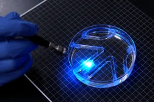
To demonstrate its capabilities, the researchers carefully submerged the robot in a large petri dish filled with water. Using a manual light source, they maneuvered the light over the bot. The robot, true to its design, followed the light, rhythmically flapping its fins in response. This light-guided propulsion allowed it to successfully navigate a simple maze that the team had strategically placed within the dish, showcasing its potential for controlled movement in liquid environments.
Broader Implications and the Future of Biohybrid Robotics
This pioneering work by MIT engineers represents more than just a clever robot; it signifies a profound scientific breakthrough at the intersection of biology and engineering. It pushes the boundaries of soft robotics and offers a glimpse into a future where living tissues could power a new generation of machines.
The potential applications stemming from this research are vast and transformative. In the medical field, these biohybrid micro-robots could revolutionize targeted drug delivery, navigating the intricate pathways of the human circulatory system to deliver therapeutics precisely where needed, minimizing side effects. They could also be employed in minimally invasive surgery, performing delicate tasks within the body, or assisting in diagnostics by collecting samples from hard-to-reach areas. Furthermore, the understanding gained in culturing and patterning functional muscle tissue could directly benefit tissue engineering efforts, leading to the creation of more effective grafts for muscle repair or regeneration.
From an environmental perspective, these robust yet delicate swimming robots could be deployed for micro-exploration of aquatic environments, from monitoring water quality in oceans and lakes to investigating confined or polluted waters that are inaccessible or hazardous to human divers or conventional hardware. Their ability to move efficiently through water could also make them ideal for environmental sampling or detecting contaminants.
In defense and security, the discreet nature and potential for agile movement in water could open avenues for covert surveillance, reconnaissance, or inspection in challenging aquatic terrains.
However, despite the immense promise, several challenges remain for future research. Scalability is a key consideration; transitioning from a petri dish demonstration to real-world environments will require overcoming hurdles related to size, power, and environmental robustness. The current reliance on an external light source for power and control needs to evolve towards on-board power solutions and autonomous navigation systems for true independence. The durability and lifespan of living cells in a robotic context, including nutrient supply and waste removal, are critical long-term research areas. Developing more complex control algorithms that allow for autonomous decision-making and interaction with the environment is another frontier. Finally, for in-vivo medical applications, stringent biocompatibility and safety standards must be met, alongside addressing the ethical considerations surrounding the integration of living tissue with artificial components.
This research significantly advances the field of soft robotics, inspiring new designs that leverage the unique properties of biological materials. By demonstrating the power of a single muscle cell layer, MIT continues to solidify its leadership in bio-engineering and robotics, paving the way for machines that are not only functional but also inherently adaptable, responsive, and potentially, even self-healing. The journey towards truly bio-integrated machines has just received a powerful new impetus.