The pursuit of capturing the fundamental "heartbeat" of a chemical reaction has long been a primary objective for the global scientific community. In a landmark study published recently, an international team of researchers has successfully utilized the world’s most powerful X-ray laser to observe how energy redistributes within a molecule immediately following the absorption of light. By employing the European X-ray Free-Electron Laser (European XFEL), the team revealed that individual atoms within a single molecule do not experience energy changes uniformly; rather, they act as localized witnesses, recording distinct aspects of the molecular transformation. This breakthrough provides an unprecedented, site-specific view of photochemistry, offering a granular look at the mechanisms that govern everything from biological UV protection to the efficiency of solar energy harvesting.
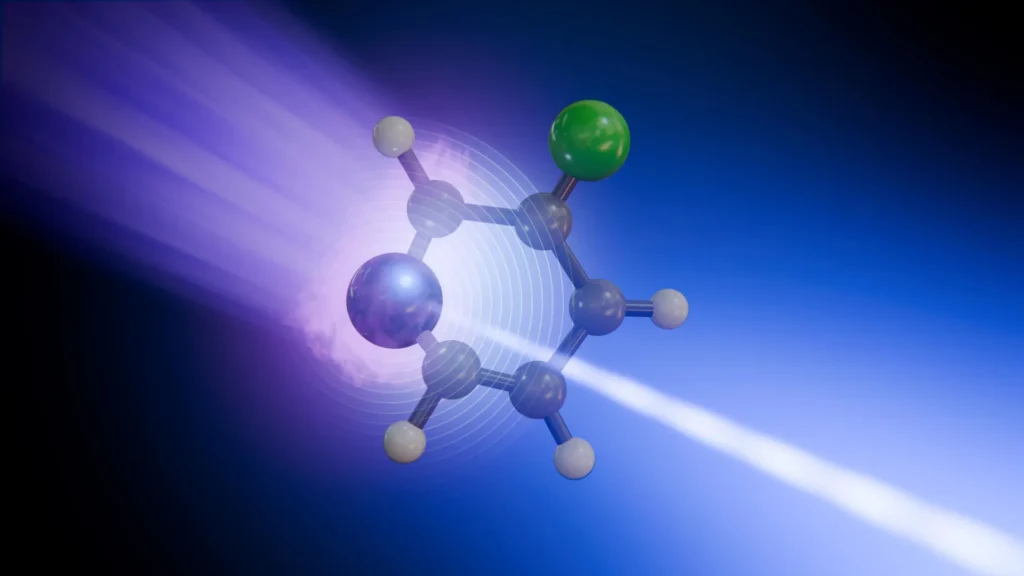
The research focused on 3-fluoropyridine, a small, ring-shaped molecule that serves as a critical model for understanding more complex biological and industrial systems. By monitoring the nitrogen and fluorine atoms within this structure, the team was able to differentiate between electronic shifts and structural vibrations, effectively deconstructing a process that occurs in trillionths of a second. This "molecular movie" marks a significant leap forward in our ability to witness the transition states of matter, which were previously considered too fast and too small to be captured in such detail.
The Mechanics of Molecular Excitation
At the center of this experiment is the phenomenon of photoexcitation. When a molecule absorbs light—such as ultraviolet (UV) radiation—its electrons are jolted into a higher energy state. This state is inherently unstable, and the molecule must find a way to dissipate this excess energy to return to its "ground state." The path it takes to achieve stability determines the outcome of the chemical process: whether the molecule remains intact, breaks apart, or undergoes a structural rearrangement.
In the case of 3-fluoropyridine, the absorption of a UV pulse causes the molecule to lose its normally flat, planar geometry. It begins to bend and twist almost instantaneously. This physical movement leads the molecule toward a "conical intersection." In the realm of quantum chemistry, a conical intersection is a vital junction where the electronic energy levels and the nuclear (atomic) positions of a molecule become inextricably linked. It acts as a high-speed funnel, allowing the electronic energy to be converted into kinetic energy—specifically, the vibration of the atoms.
Understanding how a molecule navigates this intersection is essential for predicting the stability of matter. For instance, the DNA in human cells is remarkably efficient at navigating conical intersections to dissipate UV energy as heat, preventing the chemical bonds from breaking and causing genetic mutations. The study of 3-fluoropyridine provides a simplified yet profound look at these exact dynamics.
The Role of the European XFEL and the SQS Instrument
Capturing these events requires a "shutter speed" fast enough to freeze the motion of atoms. The European XFEL, located in the Hamburg region of Germany, is one of the few facilities on Earth capable of such a feat. It generates X-ray pulses that are not only incredibly bright but also last for only a few femtoseconds (one quadrillionth of a second).
The research team utilized the Small Quantum Systems (SQS) instrument at the European XFEL to perform time-resolved X-ray photoelectron spectroscopy (tr-XPS). The experimental setup involved a "pump-probe" technique. First, a "pump" laser pulse (ultraviolet) hit the molecules to initiate the energy absorption and movement. This was followed by a "probe" pulse—a soft X-ray flash—at precisely timed intervals.
The X-ray pulse works by knocking deeply bound "core" electrons out of specific atoms within the molecule. By measuring the kinetic energy of these released electrons, the researchers could determine the chemical environment of the atom at that exact moment. Because nitrogen and fluorine atoms require different X-ray energies to release their core electrons, the researchers could "tune" their observations to look at one atom at a time, even though they were part of the same molecule.
A Tale of Two Atoms: Nitrogen vs. Fluorine
The most striking finding of the study was the divergence in the "stories" told by the nitrogen and fluorine atoms. While they are part of the same molecular ring, their responses to the energy pulse were radically different.
The fluorine atom acted as a stable observer of the molecule’s overall health. As the molecule returned to its ground state and began to vibrate, the signal from the fluorine atom tracked these vibrations with high clarity. It provided a roadmap of how the energy was cooling down into heat throughout the molecular frame.
In contrast, the nitrogen atom provided a much more complex narrative. Because the nitrogen atom is more directly involved in the initial electronic excitation of the 3-fluoropyridine ring, its signal was sensitive to both the movement of the electrons (charge density) and the changing shape of the molecule. The nitrogen atom "reported" on the chaotic period when the molecule was passing through the conical intersection, where the distinction between electronic and structural motion is blurred.
Antonio Picón, a co-author from the Instituto de Ciencia de Materiales de Madrid (ICMM-CSIC), emphasized the importance of this multi-site sensitivity. "We can now see that not every atomic site tells the same story," Picón noted. "Some atoms report where the charge is going, while others reveal how the whole molecule vibrates." This ability to decouple different types of motion within a single reaction is a major technical milestone.
Chronology of the Transformation
The entire process observed by the researchers took place within a window of approximately two picoseconds (two trillionths of a second). To reconstruct this timeline, the team had to repeat the experiment thousands of times, slightly increasing the delay between the UV "pump" and the X-ray "probe" each time.
- Time Zero: The UV laser pulse hits the 3-fluoropyridine molecule, exciting its electrons.
- 0 to 100 Femtoseconds: The molecule begins to deform from its flat shape. The nitrogen atom shows a rapid shift in its electronic environment.
- 100 to 500 Femtoseconds: The molecule reaches the conical intersection. The electronic energy begins its rapid conversion into structural vibrations. The signals from the nitrogen atom are at their most complex during this phase.
- 500 Femtoseconds to 1 Picosecond: The molecule settles back into its electronic ground state, but it is now "hot"—it is vibrating violently.
- 1 to 2 Picoseconds: The fluorine atom provides a clear signal of these vibrations as they begin to redistribute across the molecule and eventually dissipate.
Supporting Data and Theoretical Integration
The raw data from the X-ray flashes would be nearly impossible to interpret without the aid of advanced computational chemistry. To make sense of the electron energies recorded at the SQS instrument, the team employed sophisticated computer simulations. These models allowed them to visualize the "potential energy surfaces" the molecule was traveling along.
By comparing the experimental signals to the theoretical models, the researchers confirmed that the changes in the X-ray spectra directly corresponded to specific bond angles and electron densities. This synergy between "big science" hardware (the XFEL) and "big data" software (quantum simulations) is increasingly becoming the standard for modern chemical physics.
Broader Implications: From DNA to Solar Cells
While 3-fluoropyridine is a relatively simple molecule, the implications of being able to watch its energy flow are vast. The study serves as a proof-of-concept for investigating much larger and more consequential systems.
1. Biological Protection and DNA:
Understanding how nitrogen-containing rings (like those found in DNA bases) handle UV light is fundamental to biology. If we can map exactly how energy moves through these rings to prevent damage, we may gain new insights into the origins of genetic stability and the mechanisms of skin cancer at a molecular level.
2. Next-Generation Photovoltaics:
In the field of renewable energy, scientists are constantly looking for materials that can harvest light and convert it into electricity with minimal loss. By observing how energy "leaks" or gets trapped as vibrations in synthetic molecules, engineers can design more efficient solar cells and light-activated catalysts.
3. Drug Design and Photocatalysis:
Many modern medicines are synthesized using light-driven reactions, which are often cleaner and more specific than traditional heat-based chemistry. Real-time observation of these reactions allows chemists to "see" the intermediate steps, enabling them to optimize the production of complex pharmaceuticals.
Official Responses and the Future of the Field
The success of the experiment has been hailed as a validation of the European XFEL’s mission. Daniel Rivas, a guest scientist at SQS and co-author of the study, remarked on the facility’s unique capabilities. "This is what European XFEL was built to enable: watching chemical change where it begins, at specific atomic sites and on its natural timescale," Rivas said. He added that the combination of "multi-site sensitivity" and "femtosecond resolution" is opening a new window into the microscopic mechanisms of the universe.
The research also highlights the power of international collaboration. The study involved scientists from Germany, Spain, and several other nations, bringing together experts in laser physics, molecular chemistry, and theoretical modeling.
Looking forward, the team plans to apply this technique to more complex "heterostructures"—molecules made of different types of building blocks where the flow of energy is even more intricate. As X-ray laser technology continues to evolve, the "shutter speed" will become even faster, and the sensitivity even higher, potentially allowing scientists to watch the motion of individual electrons in real time.
In conclusion, the ability to use atoms as "local reporters" within a molecule represents a paradigm shift in chemistry. We are moving away from a time when we could only see the starting materials and the end products of a reaction. Now, we are standing in the middle of the reaction itself, watching the energy flow, the bonds bend, and the atoms vibrate in a choreographed dance of physics and chemistry. This new level of detail is not just a scientific curiosity; it is the foundation upon which the next century of materials science and biotechnology will be built.