In a significant stride toward revolutionizing cancer diagnostics and personalized medicine, researchers led by MIT Professor Kripa Varanasi, in collaboration with colleagues at MIT and Johns Hopkins University, have unveiled a groundbreaking handheld device designed to gently collect living cells from precise locations. This innovation holds immense promise for the earlier and more effective detection of ovarian cancer, a disease notoriously difficult to diagnose in its treatable stages, and could pave the way for highly individualized treatment strategies. The new technology addresses a critical unmet need in oncology, particularly for cancers where early detection dramatically impacts survival rates.
The Silent Scourge: Understanding Ovarian Cancer’s Lethality
Ovarian cancer stands as one of the deadliest forms of cancer affecting women, largely due to the timing of its diagnosis. The stark reality is that when detected early—at stages 1 or 2—the five-year survival rate can exceed 90 percent. However, the vast majority of cases (approximately 70-75%) are diagnosed at advanced stages (3 or 4), when the cancer has already spread beyond the ovaries, plummeting the five-year survival rate to less than half that figure, often in the range of 20-30%. This dramatic disparity underscores the urgent need for more effective early detection methods. The insidious nature of ovarian cancer stems from its vague symptoms, which often mimic more benign conditions like digestive issues or menstrual discomfort, leading to misdiagnosis or delayed investigation. There is currently no reliable, population-wide screening test for ovarian cancer, unlike mammograms for breast cancer or Pap smears for cervical cancer, further contributing to late-stage diagnoses.
A Paradigm Shift: The Fallopian Tube Connection
For decades, ovarian cancer research primarily focused on the ovaries themselves as the origin point of the disease. However, about 20 years ago, a pivotal discovery began to reshape this understanding. Researchers identified that many cases of high-grade serous ovarian cancer (HGSOC), which accounts for 70-80% of all ovarian cancers and is the most common and aggressive type, actually originate not in the ovaries, but in the fimbriated ends of the fallopian tubes. These fimbriae are finger-like projections at the end of the fallopian tube that sweep over the ovary to pick up an egg. It is in these areas that precursor lesions, known as serous tubal intraepithelial carcinomas (STICs), often develop.
This paradigm shift opened new avenues for both prevention and early detection. For women at increased risk of ovarian cancer, particularly those with BRCA gene mutations, opportunistic salpingectomy—the removal of the fallopian tubes while preserving the ovaries—has emerged as a viable prevention strategy after childbearing years. This procedure largely maintains normal hormone production, offering a significant benefit over traditional oophorectomy (ovary removal). Despite this crucial insight, detecting these precursor lesions remains incredibly challenging in a clinical setting. The STICs can be microscopic, often just a few cells thick, making them exceedingly difficult to identify through imaging techniques or to sample accurately without invasive procedures. The existing diagnostic gold standard involves surgically removing suspicious tissue, followed by a time-consuming and destructive process of chemical preservation and sectioning for microscopic examination by a pathologist. This method, while effective for definitive diagnosis, renders the cells non-viable, preventing crucial live cell studies.
Innovation at the Interface: Engineering a New Diagnostic Frontier
The critical need for a non-destructive, targeted cell collection method inspired the interdisciplinary team at MIT and Johns Hopkins. Professor Kripa Varanasi, the Maher A. Elmasri Professor of Mechanical Engineering at MIT, whose lab specializes in fluid-surface interfaces, spearheaded the engineering effort. He collaborated with Dr. Rebecca Stone, a gynecologic oncologist and the Stoddard and O’Neil Professor in Gynecologic Oncology at Johns Hopkins University School of Medicine, and Professor Angela Belcher, MIT’s James Mason Crafts Professor and a professor of biological engineering and of materials science and engineering. This powerful collaboration was catalyzed by funding from Break Through Cancer, a foundation dedicated to fostering interdisciplinary teams to tackle challenging cancer problems. Their collective project specifically aimed at developing novel approaches for the early detection of ovarian cancer, with this cell-collection technology forming a cornerstone of that broader initiative.
The initial challenge was clear: how to collect living cells from specific, potentially precancerous regions of the fallopian tube without damaging the surrounding tissue or compromising cell viability. Traditional methods, as Varanasi noted, are "very time-consuming and destructive to the cells." The MIT researchers witnessed firsthand the limitations of current surgical and pathological workflows during visits to operating rooms at Johns Hopkins. This exposure provided a crucial spark. Varanasi recalled, "We do a lot of work on fluid-surface interfaces in my lab, and we realized we could use a fluid instead of a scalpel or brush, because when you flow a fluid it applies shear stress at the interface." The idea was bolstered by surgeons’ observations that some cells in specific anatomical locations were loosely adherent and would detach during routine washing procedures. This insight formed the basis for a novel, non-invasive cell collection strategy.
Dr. Stone emphasized the synergistic nature of this collaboration: "This is exactly the kind of problem that benefits from bringing clinicians and engineers together. We understand the clinical need, while the MIT team brings a very different perspective from fluid mechanics and engineering. That combination allowed us to approach the problem in a new way." This convergence of clinical expertise and engineering ingenuity was pivotal in conceptualizing a device that could overcome long-standing diagnostic hurdles.
Precision Sampling: How the Microfluidic Device Works
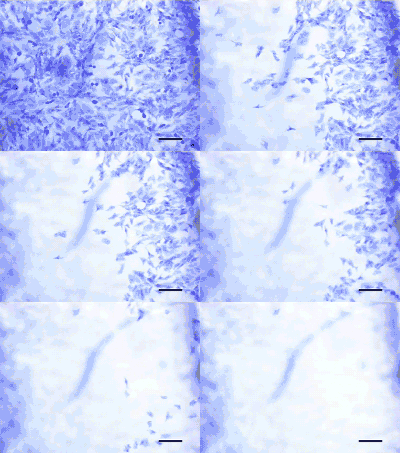
The result of this collaborative endeavor is an innovative, handheld microfluidic device, fabricated using advanced 3D printing techniques. The device operates on a sophisticated principle of controlled fluid dynamics to gently detach cells. It forms a vacuum seal with the tissue, localizing the fluid flow to a tiny, precise region. Two syringes are integral to its operation: one creates the vacuum to firmly hold the device against the tissue, ensuring no leakage, while the second syringe pushes a carefully controlled volume of liquid through the microfluidic channel. This flowing liquid generates a tunable shear stress parallel to the tissue surface, which gently and non-destructively dislodges living cells.
Bert Vandereydt, an MIT postdoc and co-first author of the study published in the journal Device, elaborated on the mechanism: "We came up with this device where one syringe creates a vacuum that holds it against the tissue, and a second syringe pushes liquid through it. The vacuum creates a seal, so nothing leaks, and then we locally apply what is basically a microfluidic chip on the tissue that gently shears the cells off." This method stands in stark contrast to conventional biopsy techniques that involve cutting or scraping tissue, which often damage cells and yield a mix of viable and non-viable material. The ability to control the shear stress applied to the tissue is a significant advantage, allowing researchers to optimize cell detachment while preserving cell viability. The study demonstrated that cells collected using this technique remained highly viable and proliferated in culture much more readily than those obtained through traditional methods.
From Bench to Bedside: Validating the Technology with Human Samples
To validate the device’s efficacy and clinical relevance, the researchers embarked on a rigorous testing phase using fresh human fallopian tube samples. This involved a demanding logistical dance, with the MIT team needing to be on call for immediate sample shipments from their collaborators at Johns Hopkins. "The samples could come at any time. Sometimes, we’d get an email from our collaborators at 11 p.m. saying ‘There are two fallopian tubes coming tomorrow,’" Vandereydt recounted. This round-the-clock dedication was essential to ensure the samples were processed while the cells were still viable.
The successful collection of living cells from these human fallopian tubes represented a major breakthrough. Crucially, the researchers were able to cultivate these collected cells into organoids—miniature, self-organizing 3D tissue cultures that mimic the structure and function of the original organ. Organoids are invaluable tools for disease modeling, allowing scientists to study cancer progression in a highly realistic environment outside the body. They also provide a platform for testing the efficacy of different drugs on a patient’s specific cancer cells, moving closer to true personalized medicine. After the experiments, the tissue samples were sent back to Johns Hopkins for conventional histopathological analysis, allowing for a direct comparison and validation of the device’s targeted sampling capabilities.
Professor Belcher highlighted the synergistic potential: "We are developing optical approaches to identify suspicious regions of tissue, and this technology could allow us to collect living cells from exactly those locations. Being able to first see where the disease may be emerging and then collect those cells for further study could be very powerful." This vision of combining advanced imaging with precision cell collection offers a comprehensive strategy for early detection and characterization of nascent cancers.
Beyond Ovarian Cancer: A Versatile Tool for Oncology
While the initial focus of this research was ovarian cancer, the fundamental principle of the device—the gentle, targeted collection of living cells through tunable shear stress—makes it remarkably versatile. The researchers tested the device on various cell types and discovered that the approach is "agnostic to the disease," as Vandereydt put it. By adjusting the level of shear stress, the device can effectively collect cells with different adhesion properties. For instance, very loosely adherent prostate cancer cells might detach at a low stress of 1 pascal, while more strongly adherent bone cancer cells might require up to 5 pascals of stress for efficient collection. This adaptability implies that the device could be applied to a wide array of cancers where obtaining viable cells from specific regions is critical for diagnosis, research, and treatment development. This broad applicability significantly amplifies its potential impact across the field of oncology.
Transforming Cancer Care: Implications for Diagnostics and Personalized Medicine
The implications of this technology are far-reaching, promising to transform several aspects of cancer care.
1. Revolutionizing Pathology: The ability to collect living cells from specific areas while preserving the surrounding tissue for traditional histopathology is a game-changer. Dr. Stone envisions integrating this technology into routine histopathology workflows, creating "a powerful new way to study carcinogenesis and fundamental biology directly from human tissue." Pathologists could not only examine tissue architecture but also immediately access living cells for functional studies, offering a more holistic view of the disease.
2. Advancing Personalized Medicine: One of the most exciting prospects is the acceleration of personalized medicine. By growing patient-derived organoids from the collected living cells, clinicians could test how an individual patient’s specific cancer cells respond to various chemotherapies, immunotherapies, or targeted drugs ex vivo. This "pre-clinical trial" for each patient could guide treatment decisions, minimizing ineffective therapies and optimizing outcomes, thereby moving towards truly precision oncology. This approach holds the potential to dramatically improve treatment efficacy and reduce side effects by avoiding trial-and-error treatments.
3. Enhancing Early Detection Strategies: While the initial application of the device will likely involve tissue already removed from the body—an easier pathway for regulatory approval—the long-term vision includes using it for in vivo sampling. This could involve integrating the device into an endoscope or a minimally invasive surgical tool, allowing clinicians to collect samples from suspicious regions inside a patient’s body without requiring extensive surgery. Such a capability would represent a monumental leap forward in early cancer detection, particularly for hard-to-reach cancers like ovarian, pancreatic, or lung cancer, where early diagnosis is currently a major challenge.
4. Deepening Disease Understanding: Access to living cells from precancerous lesions or early-stage tumors will provide unprecedented opportunities for researchers to study the fundamental biology of cancer initiation and progression. Understanding the molecular and cellular changes that occur as healthy cells transform into cancerous ones is crucial for developing new preventive strategies and therapeutic targets.
The Road Ahead: Challenges and Aspirations
The journey from a promising research device to a widely adopted clinical tool involves navigating complex regulatory pathways, particularly for in vivo applications. The researchers’ strategy of initially targeting excised tissue makes regulatory approval more attainable, laying the groundwork for broader clinical use. Continued research will focus on miniaturization, integration with imaging technologies, and further validation across diverse cancer types.
Professor Varanasi expressed his profound motivation for this work: "If this work can ultimately help women by enabling earlier detection of ovarian cancer, I would find that incredibly fulfilling. That is really what motivates us – taking the science and engineering we develop in the lab and using it to make a difference in people’s lives." The support from the Break Through Cancer Foundation has been instrumental, fostering an environment of collaborative innovation that brought together experts from various disciplines to tackle the multifaceted challenges of cancer.
This new handheld device represents a powerful testament to the impact of interdisciplinary scientific collaboration. By bridging the fields of mechanical engineering, biological engineering, and gynecologic oncology, the team has created a technology with the potential to transform the landscape of cancer diagnosis, treatment, and research, offering a new beacon of hope for patients facing some of the most challenging forms of the disease.