A pivotal advancement in microscopic imaging, reminiscent of a century-old breakthrough that transformed light microscopy, has now been achieved in the realm of electron microscopy. Physicists at the University of California, Berkeley, have developed a novel "laser phase plate" that promises to dramatically enhance the capabilities of cryoelectron microscopy (cryo-EM) and cryoelectron tomography (cryo-ET), opening unprecedented windows into the intricate world of biological molecules, particularly those previously too small or elusive to observe in detail. This innovation, published in the prestigious journal Science, is poised to accelerate drug discovery, deepen our understanding of disease mechanisms, and fundamentally reshape structural biology.
Echoes of a Nobel-Winning Past: The Legacy of Phase Contrast
Almost a century ago, in 1930, Dutch physicist Frits Zernike made a discovery that revolutionized the study of living cells. He realized that when light passes through biological material, it doesn’t just get absorbed or scattered, causing changes in brightness; it also experiences subtle shifts in its phase, or the timing of its wave peaks. While the human eye cannot directly perceive these phase shifts, Zernike ingeniously devised a method to convert them into visible differences in brightness. By manipulating the phase of the unscattered light by 90 degrees, he made previously faint or nearly invisible cellular structures, such as mitochondria and nuclei, stand out with remarkable clarity against their transparent, water-rich backgrounds. This technique, known as phase-contrast microscopy, eliminated the need for chemical stains, which invariably killed the cells, thereby allowing scientists to study dynamic processes in living specimens for the first time. Its profound importance in cell biology was formally recognized when Zernike was awarded the Nobel Prize in Physics in 1953. This foundational principle, enabling enhanced contrast without damaging the sample, laid the groundwork for countless discoveries and became an indispensable tool in laboratories worldwide.
The Electron Microscope’s Journey: From Concept to Cryo-EM Revolution
The advent of the electron microscope in the 1930s by Ernst Ruska and Max Knoll offered an exponential leap in magnification and resolution compared to its optical counterpart. Utilizing beams of electrons instead of light, electron microscopes could magnify objects roughly 10,000 times more than conventional light microscopes, revealing structures at the nanoscale, including individual proteins. However, applying Zernike’s phase-contrast principle to electron microscopy proved far more challenging. Early attempts encountered significant technical hurdles; devices designed to alter the phase of electron beams often introduced image instability, reduced resolution, or significantly weakened the electron beam itself, negating the potential benefits.
For decades, scientists grappled with how to visualize biological samples effectively under electron microscopes. Electron beams, while offering superior resolution, are also highly energetic and can cause severe radiation damage to delicate biological molecules. Traditional preparation methods, such as dehydration and heavy metal staining, often distorted or destroyed the native structures researchers sought to study. This changed dramatically with the development of cryoelectron microscopy. Pioneering work in the 1960s and 70s, notably by Joachim Frank, Richard Henderson, and Jacques Dubochet, alongside significant contributions from UC Berkeley’s Robert Glaeser, led to a method where biological samples could be rapidly frozen in a thin layer of vitreous (glass-like) ice. This vitrification process preserves molecules in their near-native state, protecting them from radiation damage and allowing for imaging without stains or fixation.
Glaeser, now a UC Berkeley professor emeritus of molecular and cell biology, was instrumental in demonstrating that cooling specimens helped preserve them during imaging and proposed using a less intense electron beam to examine thousands of frozen molecules at once, further limiting damage. By combining images from multiple angles and computationally reconstructing 3D structures, cryo-EM became a powerful tool for determining the structures of large biomolecules, sidestepping the often-insurmountable challenge of crystallizing proteins required for X-ray crystallography. This transformative technique was recognized with the 2017 Nobel Prize in Chemistry, an award that specifically acknowledged Glaeser’s foundational contributions. While cryo-EM has revolutionized structural biology and significantly sped up drug discovery by revealing the precise architecture of proteins and viruses, it still faces a critical limitation: most human proteins, particularly smaller ones, remain exceedingly difficult to image with sufficient clarity due to inherent signal-to-noise limitations.
Unveiling the Unseen: The Laser Phase Plate Technology
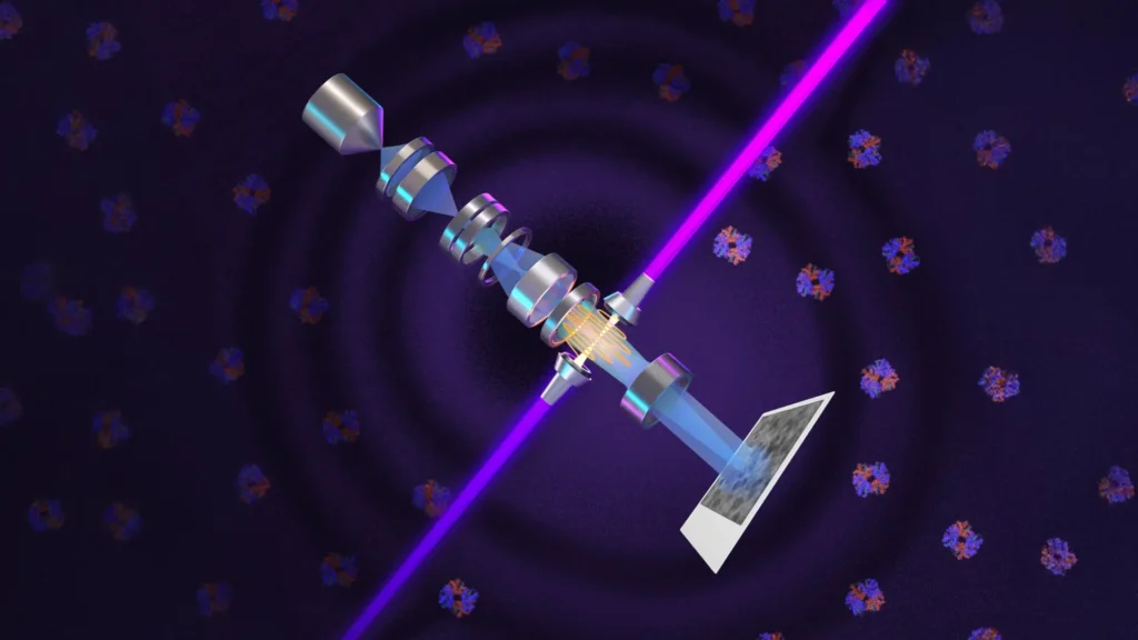
The breakthrough by Holger Müller, a UC Berkeley professor of physics and faculty scientist at Lawrence Berkeley National Laboratory, and his team directly addresses this persistent challenge by bringing the long-sought phase-contrast enhancement to electron microscopy. Their device, dubbed a "laser phase plate," functions by interacting directly with the microscope’s electron beam, precisely altering its phase to dramatically increase image contrast. This elegant solution, first theoretically proposed by Müller and Glaeser in 2010, offers a way to improve contrast without sacrificing electron beam intensity or damaging the specimen.
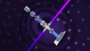
At the heart of this innovation is what researchers describe as the world’s most intense focused continuous-wave laser. Unlike conventional methods that simply illuminate a specimen, this extraordinary laser, operating at approximately 75 kilowatts of power, is confined within a spherical chamber lined with highly reflective mirrors. The light repeatedly reflects through this cavity more than 10,000 times, dramatically amplifying its intensity and focusing the beam into an incredibly tiny spot—just a few microns in size. This immense power, comparable to that used in industrial welding or military-grade lasers, is precisely what’s needed to induce the necessary 90-degree phase shift in the electron beam as it passes through. This adjustment makes it significantly easier to distinguish small molecules, such as hemoglobin, from their surrounding environment, and to visualize intricate structures within cells, like the nucleus and mitochondria, with unprecedented clarity.
Müller named his advanced cryo-EM instrument, equipped with the new laser phase plate, "Theia," after the ancient Greek Titaness associated with light and radiance. He describes Theia as the "Formula 1 microscope," noting that its enhanced electron optics already provide better resolution than standard cryo-EM instruments, even before the laser’s activation. With the laser phase plate, he anticipates it will become "the world’s best instrument overall."
In a parallel and complementary effort, Bridget Carragher, founding technical director of imaging at Biohub in Redwood City, California, is directing the construction of a related microscope. Building on theoretical research by Müller and his collaborators, Carragher’s team is developing an instrument that utilizes two lasers rather than one. Arranged at right angles to each other, each laser operates at approximately half the power of Müller’s single-laser system. This dual-laser configuration is designed to prevent components from burning out while also minimizing optical distortions that could compromise image quality. Both research teams are collaborating closely with Thermo Fisher Scientific, a leading manufacturer of cryo-EM instruments, indicating a clear path toward broader adoption and potential commercialization of this groundbreaking technology.
Breaking the Size Barrier: Imaging the Human Proteome
One of the most profound impacts of the laser phase plate technology is its ability to overcome the longstanding "size barrier" in cryo-EM. Conventional cryo-EM systems struggle to reliably image proteins smaller than approximately 70 kilodaltons (kDa). This limitation is particularly significant because proteins below this threshold account for an estimated 90% of the human proteome—the complete collection of proteins produced by the human body. Many crucial proteins involved in signaling pathways, disease progression, and fundamental cellular processes fall within this "invisible" range.
With the laser phase plate, researchers have already demonstrated the ability to image proteins as small as 50 kilodaltons, a remarkable leap forward. The Berkeley team showcased this capability by reconstructing images of two proteins: aldolase, a relatively large muscle protein that modern cryo-EM can already image with ease, and hemoglobin, a much smaller protein responsible for oxygen transport in the bloodstream, which lies near the lower size limit for conventional cryo-EM. The improvement in resolution was evident for both, but particularly pronounced for hemoglobin, unequivocally demonstrating the technology’s potential for smaller molecules. "For the most challenging cases – small particles, bad specimens – the laser produces a very considerable advantage," Müller noted. He further emphasized, "This could fill an enormous gap in our knowledge of protein structures that can’t be crystallized or are too small for today’s cryo-EM."
Looking ahead, Müller believes the technology could eventually enable scientists to study proteins as small as 17 kilodaltons, roughly the size of myoglobin, a critical oxygen-binding protein in muscle tissue. Achieving this ambitious goal may involve further refinements, such as using a tightly focused electron beam instead of the defocused beam currently required to produce sufficient contrast without the laser phase plate. Müller posits that the laser system should make it possible to obtain contrast directly from phase changes in a focused electron beam. If successful, this adjustment could potentially double the contrast and signal-to-noise ratio once again, amplifying the already significant improvements delivered by the laser phase plate.
Beyond Static Structures: The Promise of Cryo-Electron Tomography (Cryo-ET)
While cryo-EM excels at determining the structures of isolated macromolecules, an even greater impact of the new laser technology is anticipated in cryo-electron tomography (cryo-ET). Cryo-ET is a newer, more advanced approach that combines images captured from multiple angles to construct detailed three-dimensional views of proteins and other molecules within their natural cellular surroundings. Unlike techniques that study isolated molecules in a solution, cryo-ET reveals how proteins are arranged, interact, and function within the crowded, dynamic environment of a cell.
As Bridget Carragher aptly describes, "With cryo-ET, we’re looking at small, very complicated cellular material that’s incredibly crowded inside the cell. It’s like a forest of trees, and you’re trying to find one leaf on one tree in there. Cryo-ET needs a dramatic step forward in contrast, so we can start to see what’s going on inside the cell. That’s what the laser phase plate promises to give us." By significantly enhancing the signal-to-noise ratio and contrast, the laser phase plate will empower cryo-ET to visualize the complex interplay of molecular machines in situ, providing unparalleled insights into cellular processes that were previously obscured by low contrast and resolution. This ability to observe proteins in their functional context is a game-changer for understanding fundamental biology and disease mechanisms.
A Decade and a Half of Innovation: The Road to "Theia"
The journey from a theoretical concept to a working technological marvel spanned 15 years for Holger Müller and his team. Following their 2010 proposal, a significant portion of the early research was supported by the National Institutes of Health. The initial major challenge was the extraordinary feat of generating and confining an intense laser beam within a minuscule area. It took a decade of dedicated engineering and experimentation to develop the intricate system of mirrors and the spherical chamber that could build up the "brightest continuous laser focus ever," capable of inducing the necessary 90-degree phase shift in the electron beam.
The team first validated their concept by installing a prototype laser phase plate in one of Glaeser’s older electron microscopes, proving the viability of the approach. Subsequent financial support from Biohub was crucial, enabling the acquisition of a customized, state-of-the-art Thermo Fisher Krios cryo-EM microscope, which was then meticulously modified to integrate the sophisticated laser technology, culminating in the creation of Theia.
In their latest research, the scientists rigorously tested the upgraded microscope using six diverse samples, varying in size and preparation methods. The laser consistently delivered improved image resolution, with particularly significant gains for specimens that pose considerable challenges for standard imaging techniques. This systematic validation underscores the robust and broadly applicable nature of the laser phase plate. The Berkeley team’s comprehensive findings were published in the journal Science, while details of Biohub’s two-laser design are available in a recently posted preprint, highlighting the rapid pace of development and the collaborative spirit within the scientific community.
Implications for Science and Medicine: Accelerating Discovery
The implications of the laser phase plate technology extend far beyond the confines of academic research, promising to catalyze advancements across numerous scientific and medical disciplines. By enabling clearer, higher-resolution imaging of smaller proteins and molecular machines within their native cellular environments, this breakthrough stands to significantly accelerate drug discovery. Pharmaceutical researchers will be able to visualize drug targets—often small proteins—with unprecedented detail, facilitating the rational design of new therapeutic compounds and understanding how existing drugs interact with their targets at the molecular level. This could drastically reduce the time and cost associated with developing new treatments for a wide range of diseases.
As Stephani Otte, Biohub’s Vice President of Imaging Science, eloquently states, "This technology is a step function change for biology. We are going to be able to see how molecular machines operate inside the living cell, in context, for the first time. What was once invisible will become visible – and that changes everything about how we understand disease." This newfound clarity will be particularly impactful in areas like neurodegenerative diseases (e.g., Alzheimer’s, Parkinson’s), cancer, and infectious diseases, where the precise structural and dynamic interactions of proteins are critical to understanding disease progression and developing effective interventions. For example, visualizing the assembly of viral proteins or the aberrant aggregation of disease-causing proteins in their cellular context could unlock new therapeutic strategies.
Furthermore, this technology will open entirely new avenues of basic research, allowing scientists to explore the architecture and function of the vast majority of the human proteome that has remained structurally uncharacterized due to size limitations. It will provide unparalleled insights into fundamental biological processes, from protein folding and signaling to cellular trafficking and metabolism, paving the way for a deeper, more holistic understanding of life at the molecular scale. The close collaboration with Thermo Fisher Scientific also signals the potential for widespread adoption of this technology, indicating that these advanced microscopy capabilities may soon become more accessible to the broader scientific community, further amplifying its impact.
The Berkeley research team behind this monumental achievement includes Robert Glaeser; postdoctoral fellows and co-first authors Petar Petrov and Jessie Zhang; staff scientist Jonathan Remis; postdoctoral fellow Hang Cheng; and current and former physics graduate students Jeremy Axelrod, Eric Cooper, Ian Hicklin, Shahar Sandhaus, and Cooper Schnurr. At Biohub, Bridget Carragher and David Agard, the organization’s founding scientific director of imaging, jointly lead the Dynamic Structural Cell Biology group and are co-corresponding authors of the Biohub preprint, alongside Biohub engineer Pavel Olshin. Their combined efforts have ushered in a new era for biological imaging, where the previously unseen will finally become visible, transforming our understanding of health and disease.