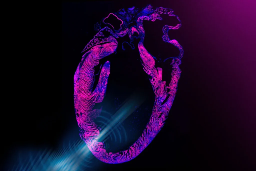
Cambridge, MA – A team of engineers at the Massachusetts Institute of Technology (MIT) has unveiled a revolutionary non-invasive pacemaker that employs ultrasound to stimulate the heart, marking a significant stride toward a surgery-free alternative to traditional cardiac implants. This innovative device, designed as a discreet chest sticker, holds the promise of transforming cardiac rhythm management by mitigating the risks associated with invasive surgical procedures. The breakthrough, detailed in a study published today in the prestigious journal Nature Biomedical Engineering, combines advanced ultrasonic technology with genetic engineering, a field known as sonogenetics, to precisely control heart contractions.
The Genesis of a Non-Invasive Solution
The core of this pioneering technology lies in a small, wearable sticker embedded with tiny transducers. These transducers emit focused ultrasound pulses that safely penetrate the chest cavity to reach the heart. At a cellular level, these ultrasound waves are engineered to trigger the opening of specific ion channels within heart cells. This action allows calcium ions to flow into the cells, initiating the crucial signal that prompts a heart cell to contract and beat. A key innovation in this design is the amplification of this effect through genetic engineering, making the heart cells exceptionally responsive to the ultrasonic stimuli.
In laboratory experiments, the researchers first applied ultrasound waves to human cardiac cells that had been genetically engineered. The results were compelling: the pulses effectively maintained healthy and consistent contractions in these cells. Moving beyond in vitro studies, the team then tested the ultrasound sticker on live rats. The device demonstrated remarkable efficacy, quickly, safely, and non-invasively correcting arrhythmias and restoring normal, regular heart contractions in the animal subjects. This dual success in both cellular and animal models underscores the potential viability of the technology.
The current prototype comprises the postage-stamp-sized ultrasound sticker and a compact, pocket-sized external device housing the necessary batteries and electronics. This design is a testament to the team’s ongoing work, building upon previous successes where they developed ultrasound stickers capable of imaging deep organs and tissues. The ultimate vision, as articulated by Professor Xuanhe Zhao, a professor of mechanical engineering and of civil and environmental engineering at MIT, is to integrate both imaging and stimulation capabilities into a single, comprehensive ultrasound sticker. "We believe you could one day have stickers on the body that could do long-term imaging deep in the body and also do stimulation for therapeutic effects, in a noninvasive closed-loop way," Zhao stated.
Addressing the Challenges of Conventional Pacemakers
The development of this non-invasive pacemaker is particularly significant when viewed against the backdrop of existing cardiac rhythm management solutions. Currently, an estimated 3 million adults in the United States alone rely on traditional pacemakers. These devices, while life-saving for millions, are surgically implanted into a patient’s chest, where they deliver electrical impulses to regulate heart rate. While modern pacemakers are highly advanced and generally safe, the surgical implantation process is not without risks.
Complications associated with traditional pacemaker implantation can include infection at the surgical site or within the bloodstream (affecting 1-2% of patients), lead dislodgement (where the wires connecting the pacemaker to the heart move out of place), pneumothorax (collapsed lung), and hematoma formation. Beyond the immediate surgical risks, patients face the need for battery replacements, typically every 7 to 10 years, which necessitates further invasive procedures. The presence of foreign bodies in the heart also carries a long-term risk of lead fracture or insulation failure. As Gengxi Lu, a co-corresponding author of the paper, emphasized, "Pacemakers are one of the most important and widely used human implants, and they have saved millions of lives. But they are invasive, and they make direct contact with the beating heart. The dream for many years has been noninvasive heart stimulation with ultrasound." The MIT innovation directly confronts these inherent challenges by offering a pathway to entirely circumvent surgery.
The Power of Sonogenetics: Engineering for Sound
The breakthrough’s scientific foundation rests heavily on sonogenetics, a relatively nascent field that draws inspiration from optogenetics. Optogenetics, a well-established technique, involves genetically modifying specific cells to respond to light, allowing researchers to precisely control neural activity. Similarly, sonogenetics aims to engineer cells to become highly sensitive and responsive to sound waves, including ultrasound.
While ultrasound has long been a staple in medical diagnostics for imaging internal structures due to its safe penetration capabilities, its therapeutic applications have also been expanding. Focused ultrasound, for instance, is being explored for treating neurological conditions such as Parkinson’s disease, Alzheimer’s, and certain brain disorders, by precisely targeting and modulating brain activity. Previous studies have also hinted at ultrasound’s ability to activate heart cells, though these effects were often inconsistent and weak, limiting their clinical utility.
Zhao’s team sought to overcome this limitation by significantly amplifying ultrasound’s therapeutic effects on the heart. In their sonogenetics approach, they first derived heart cells from embryonic stem cells in the lab using standard practices. They then introduced a specific genetic alteration into these cells. This manipulation led to the production of specialized ion channels that were designed to open far more readily in response to ultrasound waves. As Chen Gong, the paper’s first author, elucidated, "These channels can now ‘hear’ ultrasound better, and can open to let calcium in, which is what directly activates the cell and causes it to beat." This targeted genetic enhancement is what makes the heart cells exquisitely sensitive to the low-intensity ultrasound emitted by the sticker.
From Lab to a Future Clinical Reality
The envisioned clinical application of this ultrasound pacemaker is both innovative and ambitious. The team foresees a scenario where a patient could receive a one-time injection, akin to a vaccine, that would deliver the genetic alteration to cardiac cells, thereby boosting their sensitivity to the pacemaker’s ultrasound waves. This would constitute a form of gene therapy, a medical treatment modality that has seen growing acceptance and regulatory approval (e.g., by the FDA for conditions like sickle cell disease and spinal muscular dystrophy). "We think this step would be clinically translatable as a form of gene therapy that could enable noninvasive pacemakers," Gong explained, highlighting the feasibility within existing medical frameworks.
The physical design of the pacemaker is equally crucial. The postage-stamp-sized sticker is embedded with miniature ultrasound transducers. The adhesive component of the device utilizes a specialized hydrogel material, meticulously refined by Zhao’s group over years of research. This hydrogel is engineered for strong adhesion to skin and various materials, crucially allowing ultrasound waves to pass through without significant attenuation, ensuring efficient signal transmission to the heart. The transducers within the sticker can be finely tuned to generate ultrasound waves at specific frequencies, allowing for precise control over cardiac stimulation.
During the experiments with rats, after administering the sonogenetic solution through their tails, the researchers affixed a miniature version of the pacemaker to the animals’ chests. Upon activation, the stickers rapidly regulated the rats’ hearts. Individuals exhibiting bradycardia (slow heart rates) were brought to a normal rhythm, while those with irregular heartbeats were effectively stabilized, synchronizing with the rhythmic "ticks" of the ultrasound. "We can now use low-intensity ultrasound to open ion channels in cells to have very effective heart pacing," Gong affirmed. The team is now focused on miniaturizing the stickers further, enhancing integration, and improving stability and accuracy for long-term wear.
A Broader Vision for Bioelectronic Medicine
The implications of this research extend far beyond cardiac pacemaking. Professor Zhao envisions a future where similar sticker-based technologies could be deployed across various parts of the body for a multitude of therapeutic and diagnostic purposes. "In this paper, we demonstrated noninvasive pacemaking. However, we think this concept could be useful beyond just the heart," Zhao stated. "We believe you could one day have stickers over different parts of the body that could do long-term imaging, monitoring, and closed-loop therapeutic stimulation." This perspective points towards a transformative era of bioelectronic medicine, where smart, wearable devices could continuously monitor physiological parameters and deliver precise, non-invasive therapeutic interventions in real-time.
The Historical Arc of Cardiac Rhythm Management
The journey to non-invasive cardiac pacing has been a long one, marked by continuous innovation. The earliest external pacemakers emerged in the mid-20th century, cumbersome devices that delivered electrical shocks through the chest wall. The first fully implantable pacemaker, developed by Rune Elmqvist and Åke Senning in 1958, was a monumental achievement, though initial battery life was very short. Subsequent decades saw dramatic improvements in battery technology, miniaturization, lead design, and sophisticated algorithms for managing various arrhythmias. From single-chamber to dual-chamber and biventricular pacemakers, the technology has evolved to address complex cardiac conditions. The MIT team’s work represents another major leap, pivoting from electrical impulses delivered via implanted leads to acoustic waves transmitted externally, potentially ushering in a new era of patient comfort and safety.
Implications for Patient Care and the Healthcare Landscape
The potential implications of a clinically viable non-invasive ultrasound pacemaker are profound. For patients, it could eliminate the psychological and physical burden of surgery, reduce the risk of post-operative complications, and potentially improve quality of life. The accessibility of a sticker-based device could also simplify follow-up care and make cardiac rhythm management more widely available, particularly in regions with limited surgical infrastructure. From an economic standpoint, reducing the need for complex surgical procedures, hospital stays, and managing surgical complications could lead to significant cost savings for healthcare systems globally.
However, the path to widespread clinical adoption will involve rigorous testing and regulatory hurdles. Long-term safety and efficacy studies in larger animal models and eventually human clinical trials will be essential. Public acceptance of gene therapy, even for a localized and therapeutic purpose, will also be a factor. The development and regulatory approval process for such an integrated gene therapy and device approach is likely to be complex and protracted, requiring substantial investment and collaboration between academic institutions, industry, and regulatory bodies.
The Research Team and Support
The groundbreaking research was a collaborative effort involving experts from multiple institutions. The study’s lead authors from MIT included first author Chen Gong, along with Runze Li, Won Jun Song, and former postdocs Gengxi Lu, Shucong Li, and Hsiao-Chuan Liu. Crucial contributions also came from collaborators in Professor Qifa Zhou’s group at the University of Southern California (USC), as well as researchers from Harvard University, the University of California at Los Angeles, and other groups at USC. This interdisciplinary approach, spanning mechanical engineering, biomedical engineering, and medicine, was vital to the project’s success. The work received substantial support from various funding bodies, including the National Institutes of Health (NIH), the National Science Foundation (NSF), the Department of Opthamology from Research to Prevent Blindness, and the U.S. Department of War, underscoring the broad recognition of its potential impact.
Expert Perspectives and the Road Ahead
While directly quoted reactions from external cardiologists or biomedical engineers were not provided in the original announcement, the medical community is likely to view this development with immense interest and cautious optimism. Experts would undoubtedly highlight the paradigm shift from invasive electrical stimulation to non-invasive acoustic modulation combined with genetic sensitisation. The promise of reduced infection risk and avoiding lead-related complications is particularly attractive.
The immediate next steps for the MIT team will involve further miniaturization and integration of the prototype, as well as extensive preclinical studies to demonstrate long-term safety and efficacy. Scaling up the production of the genetically engineered viral vectors for the gene therapy component, ensuring their safety and specificity, will also be a critical challenge. If successful, this technology could offer a beacon of hope for patients suffering from various cardiac arrhythmias, potentially revolutionizing how heart conditions are monitored and treated, and paving the way for a new generation of smart, wearable, and non-invasive medical devices. The vision of a closed-loop system that can both image and stimulate, adaptively responding to the body’s needs, truly represents the frontier of bioelectronic medicine.