A groundbreaking development from a consortium of Austrian research institutions promises to redefine the capabilities of electron microscopy, potentially allowing scientists to visualize structures with unparalleled clarity while significantly reducing sample damage. Researchers at TU Wien, in collaboration with teams from the University of Vienna, JKU Linz, and the University of Innsbruck, have unveiled a novel approach that integrates an electron microscope with a quantum computer. This innovative system aims to harness previously unused quantum information carried by electrons, leading to the formation of clearer images with fewer electrons, a critical advantage for studying delicate biological materials. The conceptual framework has been mathematically proven, and a prototype quantum computer electron microscope is now under construction at TU Wien.
The Quantum Leap in Microscopy: Beyond Conventional Limits
Electron microscopes have long been indispensable tools in scientific research, enabling the visualization of structures far smaller than what is discernible with ordinary light. By employing a beam of electrons instead of photons, these instruments can achieve resolutions down to the atomic scale, unveiling the intricate details of materials and biological specimens. This capability has driven advancements across diverse fields, from materials science to cell biology.
However, conventional electron microscopy operates primarily by counting electrons that interact with a sample and pass through to a detector. While remarkably effective, this method inherently overlooks a wealth of quantum information embedded within each electron’s wave function. This oversight is not merely an inefficiency; it contributes to a fundamental limitation: the need for a large number of electrons to build a high-resolution image. For many samples, particularly sensitive biological specimens like proteins, viruses, or cellular organelles, prolonged exposure to an intense electron beam can cause significant radiation damage, altering their delicate structures and compromising the integrity of the observed data.
"Today, we can image tiny details on the atomic scale," explains Philipp Haslinger from the Institute of Atomic and Subatomic Physics at TU Wien. "However, this requires a large number of electrons. And not every sample can be exposed to so many electrons without being damaged. This is often a problem, particularly when imaging biological samples such as individual proteins." For instance, in cryo-electron microscopy (cryo-EM), a technique awarded the Nobel Prize in Chemistry in 2017 for its revolutionary impact on structural biology, samples are flash-frozen to mitigate damage. Yet, even with cryo-EM, high-resolution imaging still necessitates electron doses that can cause molecular changes, leading to a trade-off between image quality and sample preservation. Typical electron doses for high-resolution cryo-EM can range from 50 to 100 electrons per square Angstrom (e-/Ų), which, despite being lower than conventional EM, still accumulates significant damage over the course of an imaging session. The challenge, therefore, lies in extracting more useful information from each individual electron, thereby reducing the total electron dose required for an accurate and high-fidelity image.
Harnessing Quantum Information Through Entanglement
The innovative solution proposed by the Austrian researchers involves a radical re-imagination of the electron microscopy process. Instead of merely counting electrons, their system aims to capture and process the inherent quantum information carried by each electron. This is achieved by integrating the electron microscope with a quantum computer, creating a hybrid system that leverages the unique properties of quantum mechanics.
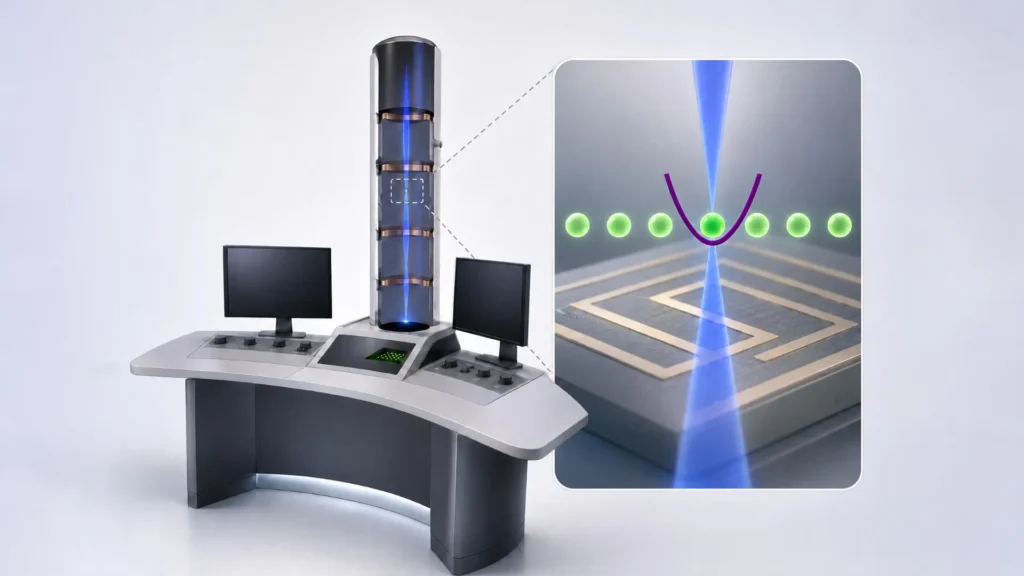
The core of their idea revolves around quantum entanglement. The researchers propose linking the electrons within the microscope to a quantum computer built around trapped ions. "Our idea is to combine the electrons with a quantum computer. We let them interact with ions that are held in place along the path of the electron beam," elaborates Elias Pescoller, first author of the publication and a doctoral student at the Institute for Theoretical Physics and the Institute of Atomic and Subatomic Physics at TU Wien. "This can, for example, create quantum entanglement between the electron and the quantum computer. The electron and the ion then share a joint quantum state."
Quantum entanglement is a phenomenon where two or more quantum particles become inextricably linked, such that the quantum state of one particle instantly influences the state of the others, regardless of the distance separating them. In this setup, as an electron passes through the sample and then interacts with an ion within the quantum computer, it can become entangled with that ion. This entanglement allows information about the electron – beyond just its presence or absence – to be encoded and stored within the quantum state of the ion. This could include subtle details about the electron’s phase, momentum, or spin that are typically lost or ignored in classical detection methods.
The Role of Quantum Computing: Amplifying Weak Signals
The true power of this approach emerges from the quantum computer’s ability to process this stored quantum information. After one electron interacts with a trapped ion and establishes entanglement, subsequent electrons can pass through and become entangled with the same or other ions in the quantum computer. By repeatedly performing carefully designed quantum operations, the system can coherently combine information from multiple electrons.
"If we perform very specific quantum-computing operations each time, we can optimally combine the information from several electrons so that we obtain a signal of maximum strength even though we use only a relatively small number of electrons," states Dennis Rätzel from the Institute of Atomic and Subatomic Physics at TU Wien. These sophisticated algorithms, crucial for translating weak quantum signals into useful imaging data, were developed in collaboration with Johannes Kofler’s team at JKU Linz.
While the fundamental imaging process still relies on electrons interacting with the sample, similar to a conventional electron microscope, the quantum computer acts as an intelligent processor. It can extract details that would otherwise be indistinguishable from random noise in classical systems. "The electrons themselves are used to image small objects, just as in any other electron microscope. But by processing the quantum information carried by these electrons in a quantum computer, we can extract significantly more information from the process," says Iva Bezinová from the Institute for Theoretical Physics at TU Wien. "What would previously have been indistinguishable from random noise can thus become a clear signal." This capability allows scientists to recover valuable structural and compositional details that would be impossible to identify using ordinary electron counting alone, fundamentally overcoming the statistical limits that traditionally constrain electron microscopy.
Historical Context and Evolution of Electron Microscopy
The journey of electron microscopy began nearly a century ago, revolutionizing our ability to peer into the microscopic world. In 1931, German physicists Ernst Ruska and Max Knoll developed the first electron microscope, for which Ruska was later awarded the Nobel Prize in Physics in 1986. Their invention surpassed the theoretical resolution limits of light microscopes, which are constrained by the wavelength of visible light.
Over the decades, electron microscopy evolved dramatically. Transmission Electron Microscopes (TEMs) and Scanning Electron Microscopes (SEMs) became standard tools in research and industry. However, a persistent challenge, particularly for biological samples, remained: the vacuum environment and electron beam itself could damage and dehydrate delicate specimens, obscuring their true structures.
The advent of cryo-electron microscopy (cryo-EM) in the 1980s marked a significant turning point. By rapidly freezing biological samples in a thin layer of vitreous ice, cryo-EM allowed scientists to image biomolecules in a near-native state, largely circumventing the damage caused by dehydration. Landmark improvements in detectors and image processing algorithms further propelled cryo-EM into the forefront of structural biology, culminating in the 2017 Nobel Prize for Jacques Dubochet, Joachim Frank, and Richard Henderson for their work in developing and applying the technique. While cryo-EM dramatically reduced sample damage, it did not eliminate it entirely. The need for a relatively high electron dose to achieve atomic resolution still limits the imaging of truly fragile or dynamic structures, leaving room for further innovation. The quantum-enhanced electron microscope represents the next frontier in addressing this long-standing challenge, pushing beyond the statistical limits even cryo-EM faces.
From Mathematical Proof to a Working Microscope: The Project Timeline
The current project represents a crucial step from theoretical validation to practical implementation. The researchers have meticulously demonstrated through mathematical proofs that their novel method indeed offers significant advantages over conventional techniques, particularly in terms of information extraction and dose efficiency. This theoretical foundation laid the groundwork for the ambitious experimental phase.
The consortium is now actively engaged in constructing the first prototype of this quantum computer electron microscope. This intricate integration process is underway at TU Wien’s University Service Center for Transmission Electron Microscopy (USTEM). A key component of this system, the ion-based quantum computer, has been developed by Philipp Schindler’s team at the University of Innsbruck, highlighting the interdisciplinary nature of this endeavor. The collaboration leverages the specialized expertise present across the different Austrian universities, bringing together quantum information science, quantum computing, and advanced electron microscopy.
Major funding for this ambitious consortium, which is coordinated by the University of Vienna, comes from the Austrian Science Fund (FWF) through the Cluster of Excellence quantA, a program designed to foster cutting-edge research in quantum science. Additional crucial support has been provided by the Gordon and Betty Moore Foundation, a philanthropic organization known for its investments in scientific discovery and environmental conservation. This substantial backing underscores the perceived potential and strategic importance of this research within the global scientific community.
Implications and Future Outlook: Revolutionizing Scientific Discovery
If the quantum-enhanced electron microscope operates as expected, its implications for scientific research could be profound and far-reaching. The ability to obtain clearer images with fewer electrons holds particular significance for biological imaging. Delicate biomolecules such as individual proteins, enzymes, or viral particles, which are easily denatured or structurally altered by electron radiation, could be imaged with unprecedented fidelity. This would dramatically improve our understanding of fundamental biological processes, aid in the elucidation of complex protein structures critical for drug discovery, and provide new insights into the mechanisms of disease at the molecular level. For instance, determining the precise structure of membrane proteins, which are notoriously difficult to crystallize and image, could become more accessible, accelerating the development of new therapeutics.
Beyond biology, the technology could also benefit materials science and nanoscience. Researchers could study radiation-sensitive advanced materials, catalysts, or quantum dots without inducing structural changes during the imaging process. This would open new avenues for characterizing material defects, understanding nanoscale phenomena, and designing novel materials with tailored properties.
While the mathematical proofs are robust, the next critical challenge is to experimentally validate these benefits. The successful operation of the prototype will pave the way for a new generation of electron microscopes, fundamentally redefining how scientists interact with and understand the microscopic world. The potential to overcome statistical limits imposed by classical physics means that this technology could enable discoveries that are currently impossible, pushing the boundaries of what can be seen and understood at the nanoscale.
Expert Perspectives and Institutional Collaboration
The collaborative spirit underpinning this project is a testament to the complex and interdisciplinary nature of modern scientific breakthroughs. "It is really exciting that, within the quantA Cluster of Excellence, we can combine the expertise in quantum information, quantum computing and electron microscopy available at the different universities in Austria. This allows us to launch a unique project," remarks Thomas Juffmann from the University of Vienna, emphasizing the synergistic power of collective scientific endeavor. This unique combination of expertise, spanning theoretical physics, experimental quantum computing, and advanced microscopy, is crucial for tackling such an ambitious challenge. The quantA Cluster of Excellence serves as a vital hub, fostering the environment necessary for these diverse specializations to converge on a common goal.
The integration of quantum computing into a historically classical imaging tool represents a paradigm shift. By moving beyond mere electron counting to actively process the quantum information inherent in electron-sample interactions, the Austrian consortium is poised to unlock a new dimension of observational science. This development not only promises clearer images and better sample preservation but also underscores the growing practical applications of quantum technologies in fields previously dominated by classical physics. The scientific community eagerly anticipates the experimental validation of this pioneering work, which could usher in a new era of microscopic discovery.