In a landmark achievement for the field of quantum chemistry and condensed matter physics, an interdisciplinary research team at the University of Göttingen has successfully developed a method to image the three-dimensional wavefunction of an organic molecule. This breakthrough, published recently in the journal Nature Communications, represents a significant leap forward in our ability to observe the fundamental building blocks of matter. By combining cutting-edge photoelectron spectroscopy with highly advanced mathematical reconstruction algorithms, the researchers have managed to visualize the spatial distribution of electrons within a molecule at a resolution finer than the distance between individual atoms. This development paves the way for a new era of "quantum cinematography," where scientists may soon be able to record high-speed videos of chemical reactions as they occur on a quadrillionth-of-a-second timescale.
The nature of the quantum world has historically defied the intuitive logic of classical physics. In our everyday experience, an object occupies a specific coordinate in space at any given time. However, at the subatomic level, particles such as electrons do not behave as discrete points. Instead, they are described by a "wavefunction," a complex mathematical entity that defines the probability of finding a particle in a particular region of space or with a specific momentum. These wavefunctions, specifically known as molecular orbitals when they exist within a molecule, dictate every aspect of a substance’s identity, from its color and conductivity to its ability to bond with other molecules. Despite their central importance to science, wavefunctions have remained notoriously difficult to observe directly, often described by physicists as "mathematical ghosts" that govern reality without being visible to traditional imaging techniques.
The Challenge of Visualizing the Invisible
To understand the magnitude of the University of Göttingen’s achievement, one must first appreciate why imaging a wavefunction is so difficult. According to the principles of quantum mechanics, particularly the Heisenberg Uncertainty Principle, the act of measurement itself often alters the state of the system being observed. Furthermore, a wavefunction is a complex-valued function, meaning it possesses both an amplitude (the "size" of the wave) and a phase (the "position" in the wave’s cycle). Traditional experimental methods can typically measure the intensity or the momentum of electrons, which provides information about the amplitude, but the phase information is almost always lost during the measurement process. This is known in physics as the "phase problem."
Without the phase information, scientists cannot fully reconstruct the three-dimensional shape of the orbital. Historically, researchers have relied on massive, multi-billion-dollar research facilities known as synchrotrons to gather enough data to attempt these reconstructions. Even at these facilities, the process was arduous, requiring weeks of measurement time and producing results that were often limited to two-dimensional projections or static images of very specific, highly ordered molecular layers. The requirement for such large-scale infrastructure meant that 3D wavefunction imaging remained a niche capability, inaccessible to the broader scientific community and unsuitable for studying the rapid, dynamic changes that occur during chemical reactions.
A New Methodology: From Momentum to 3D Visualization
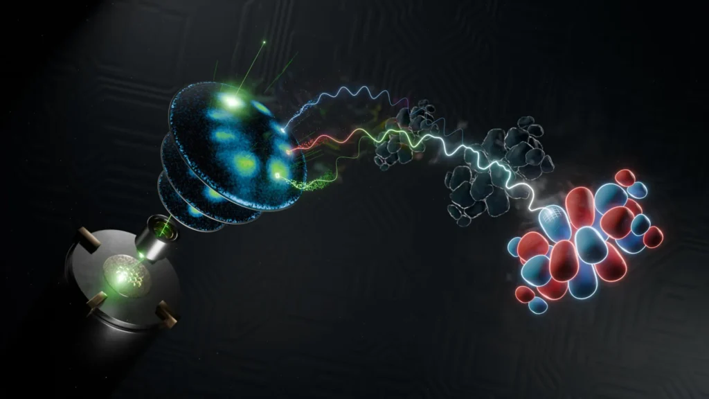
The team at Göttingen, led by Professor Stefan Mathias, Dr. Matthijs Jansen, and Dr. Wiebke Bennecke, overcame these hurdles by reimagining the relationship between experimental data and mathematical reconstruction. The team utilized a technique called photoelectron spectroscopy, which involves hitting a molecule with high-energy light—in this case, soft X-rays—to knock electrons out of their orbitals. By measuring the momentum of these "photoelectrons" as they fly away from the molecule, the researchers can gain a partial view of the wavefunction.
"The wavefunction is a fundamental quantity in quantum mechanics, yet it cannot be directly observed or measured," explained Professor Stefan Mathias. "Because we cannot measure it directly, we have to find clever ways to infer its structure from the pieces of information we can catch."
The researchers’ innovation lies in how they handled the "missing half" of the data. They developed a sophisticated suite of computer algorithms designed to solve the phase problem through an iterative reconstruction process. By taking the measured momentum data and applying physical constraints—such as the known locations of the atoms within the molecule—the algorithm can "guess" the missing phase information and refine that guess until a consistent three-dimensional image emerges.
What sets this study apart is the efficiency of the new algorithm. It requires significantly less experimental data than previous methods to produce a reliable 3D image. This efficiency allowed the team to move away from synchrotrons and instead use a laboratory-based soft X-ray source. This source produces ultrashort pulses of light, which is the key to the second major innovation of the study: the potential for high-speed imaging.
Technical Specifications and Supporting Data
The organic molecules studied by the team were only a few nanometers in size, yet the imaging technique achieved a spatial resolution that allowed researchers to distinguish features smaller than the bonds between carbon atoms. In molecular physics, carbon-carbon bonds typically measure about 0.14 nanometers. The Göttingen team’s reconstruction was precise enough to map the density of the electron cloud within these gaps, providing a vivid picture of the "molecular glue" that holds the structure together.
The laboratory-based soft X-ray source used in the experiment represents a significant technological milestone. Unlike the continuous beams often used in older spectroscopic methods, this source delivers light in femtosecond pulses. A femtosecond is one quadrillionth of a second (10^-15 seconds). To put this in perspective, a femtosecond is to one second what one second is to about 32 million years. By using such incredibly short bursts of light, the researchers have essentially created a high-speed camera shutter capable of freezing the motion of electrons.
Dr. Matthijs Jansen, co-leader of the study, emphasized the synergy between the hardware and the software: "We introduce two powerful new concepts. First, by redesigning the computer algorithm from the ground up, reliable 3D images can now be obtained using much less experimental data. Second, the experiment is based upon a powerful, lab-based soft-X-ray light source that provides ultrashort light pulses. It is the combination of these two techniques that has this remarkable impact."
Chronology of the Breakthrough
The path to this discovery has been decades in the making, following a clear chronological progression in the field of molecular imaging:
- The 1980s-1990s (The Atomic Era): The invention of the Scanning Tunneling Microscope (STM) and Atomic Force Microscope (AFM) allowed scientists to see the positions of atoms for the first time. However, these tools primarily mapped the topography of a surface rather than the internal quantum states of electrons.
- Early 2000s (The 2D Breakthrough): Researchers began using Angle-Resolved Photoemission Spectroscopy (ARPES) to map the momentum of electrons in two dimensions. This allowed for the first "orbital tomography" experiments, but they were largely restricted to flat, simple molecules on metal surfaces.
- 2010-2020 (The Synchrotron Era): Scientists moved to large-scale facilities to attempt 3D reconstructions. While successful, these experiments were "static," meaning they could only image molecules in a stable, unchanging state. The data requirements were so high that "filming" a reaction was considered a distant dream.
- 2024 (The Göttingen Milestone): The current study proves that 3D imaging can be done in a standard laboratory setting with minimal data, opening the door to time-resolved (dynamic) 3D imaging.
Broader Implications and "Stroboscopic Videography"
The most exciting implication of this research is the transition from static photography to dynamic videography. In the same way that the earliest motion pictures revolutionized our understanding of animal locomotion by freezing motion that the human eye could not track, 3D wavefunction imaging could revolutionize our understanding of chemistry.
Most chemical reactions—such as the way a plant converts sunlight into energy during photosynthesis or how a catalyst breaks down pollutants—happen on the femtosecond scale. In these moments, the molecular orbitals distort, shift, and merge to form new bonds. Until now, scientists could only observe the "before" and "after" states of a reaction.
Dr. Wiebke Bennecke, the study’s first author, believes the era of the "molecular movie" is at hand. "This technique might mean that stroboscopic videography becomes a reality, allowing us to observe not just the shape of wavefunctions, but also to see how it changes with ultrafast, even femtosecond resolution. This will mean we can learn how a molecule adapts to optical, electronic, or chemical changes and find new ways to control these interactions at the level of a few atoms."
The ability to control these interactions could lead to breakthroughs in several fields:
- Material Science: Developing new superconductors or more efficient semiconductors by understanding how electron wavefunctions behave under different temperatures or pressures.
- Pharmacology: Observing exactly how a drug molecule interacts with a protein receptor at the quantum level, allowing for the design of more precise and effective medications.
- Green Energy: Improving the efficiency of organic solar cells by visualizing how electrons move through the molecular lattice after being excited by a photon.
- Quantum Computing: Gaining better control over "qubits" by understanding the wavefunction environment that surrounds them.
Expert Analysis and Conclusion
The work of the University of Göttingen team represents a fundamental shift in experimental physics. By moving the capability of 3D quantum imaging from massive, centralized facilities into the university laboratory, they have democratized a tool that was once the preserve of a few. Furthermore, the integration of advanced algorithms to "fill in the blanks" of quantum data highlights the growing importance of computational physics in modern discovery.
While challenges remain—such as extending this technique to even more complex, non-organic molecules and further refining the speed of the X-ray pulses—the foundation has been laid. The scientific community now has a roadmap for watching the quantum world in motion. As we move closer to capturing the first true 3D "femtosecond movie" of a chemical bond breaking, the boundary between mathematical theory and observable reality continues to blur. The "mathematical ghosts" of quantum mechanics are finally being brought into the light, offering a clearer view of the universe at its most fundamental level.