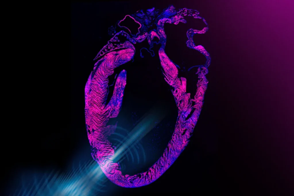
CAMBRIDGE, MA – Engineers at the Massachusetts Institute of Technology (MIT) have achieved a significant breakthrough in cardiac care, developing a novel, noninvasive pacemaker that employs ultrasound waves to stimulate the heart. This innovative design, detailed in a study published in Nature Biomedical Engineering, could one day offer a surgery-free alternative to traditional cardiac implants, revolutionizing treatment for millions living with heart rhythm disorders.
The groundbreaking device is conceived as a discreet, wearable chest sticker, featuring an array of miniature transducers. These transducers are engineered to emit precise ultrasound pulses that penetrate the chest cavity, reaching and stimulating the heart. The core mechanism involves these ultrasound waves triggering the opening of specific ion channels within heart cells. Crucially, the MIT team amplified this effect through genetic engineering, making the cardiac cells significantly more sensitive to the acoustic stimulation. When these engineered channels open, they facilitate the influx of calcium, a critical signal that prompts a heart cell to contract and beat, thereby regulating the heart’s rhythm.
In rigorous laboratory experiments, the researchers applied ultrasound waves to human cardiac cells that had been genetically engineered for enhanced sensitivity. The results were compelling: the pulses effectively maintained the cells’ healthy and consistent contractions. Further preclinical trials involved testing the ultrasound sticker on live rats. The device demonstrated its efficacy by quickly, safely, and noninvasively correcting arrhythmias and restoring normal, regular heart contractions in the animal subjects.
The team has successfully fabricated a prototype that consists of the postage-stamp-sized ultrasound sticker and a compact, pocket-sized external unit housing the necessary batteries and electronics. This development builds upon the group’s prior work, which included demonstrating a similar sticker design capable of using ultrasound for imaging deep organs and tissues. The ambitious next phase involves integrating these two functionalities – imaging and stimulation – into a single, comprehensive ultrasound sticker, enabling simultaneous monitoring and regulation of cardiac activity in a closed-loop system.
"We believe you could one day have stickers on the body that could do long-term imaging deep in the body and also do stimulation for therapeutic effects, in a noninvasive closed-loop way," stated Xuanhe Zhao, a professor of mechanical engineering and civil and environmental engineering at MIT, underscoring the broad potential of this technology.
The research findings were published today in the prestigious journal Nature Biomedical Engineering. The study’s lead authors from MIT include Chen Gong (first author), along with Runze Li, Won Jun Song, and former postdocs Gengxi Lu, Shucong Li, and Hsiao-Chuan Liu. Collaborators also include Professor Qifa Zhou’s group at the University of Southern California (USC), as well as researchers from Harvard University, the University of California at Los Angeles, and other groups at USC.
The Landscape of Cardiac Pacing: A Critical Need for Innovation
Today, approximately 3 million adults in the United States alone live with pacemakers, and this number is steadily rising globally. Traditional pacemakers are small, battery-powered devices surgically implanted into a patient’s chest, typically beneath the skin near the collarbone, with leads extending into the heart chambers. These devices deliver precise electrical impulses to regulate the heart rate, correcting bradycardia (slow heart rate) or other arrhythmias that can lead to symptoms like dizziness, fatigue, and even life-threatening cardiac events.
While implantable pacemakers represent a well-established and generally safe medical treatment that has undoubtedly saved millions of lives since their widespread adoption in the mid-20th century, they are not without their inherent risks and limitations. As Gengxi Lu, a co-corresponding author of the paper, noted, "Pacemakers are one of the most important and widely used human implants, and they have saved millions of lives. But they are invasive, and they make direct contact with the beating heart. The dream for many years has been noninvasive heart stimulation with ultrasound."
The invasive nature of traditional pacemaker implantation carries potential complications such as infection at the surgical site, lead fracture or dislodgement, venous obstruction, pneumothorax, and rare but serious cardiac perforation. Patients also face the need for periodic battery replacements, which necessitate further surgical procedures, typically every 5 to 10 years. Moreover, the presence of leads within the heart can sometimes lead to long-term issues, including tricuspid valve regurgitation or erosion of the heart tissue. For younger patients or those with active lifestyles, these considerations can significantly impact their quality of life and long-term health management. The global market for pacemakers and cardiac rhythm management devices is substantial, valued at billions of dollars, indicating the widespread reliance on these interventions and the significant opportunity for less invasive alternatives.
Sonogenetics: Harnessing Sound to Control Cellular Function
The MIT team’s innovation hinges on the sophisticated application of sonogenetics, a relatively nascent field that draws parallels with optogenetics. Optogenetics, a revolutionary neuroscience technique, involves genetically manipulating specific cells to make them responsive to light, allowing researchers to precisely control neural activity with unprecedented specificity. Similarly, sonogenetics aims to genetically engineer cells to respond to sound waves, including ultrasound.
Ultrasound, encompassing a broad range of acoustic waves, is already a cornerstone of modern medicine. It safely penetrates the body, and its waves reflect and resonate off internal structures in characteristic ways, enabling technicians to create detailed images of organs and tissues in real-time. Beyond imaging, ultrasound can be precisely directed and focused to achieve therapeutic effects, particularly in neurology, where scientists are actively exploring its use to treat debilitating conditions such as Parkinson’s disease, Alzheimer’s, and other brain disorders by modulating neural activity.
While previous studies in animals had demonstrated that focused ultrasound could safely activate heart cells, the effect was often inconsistent and weak, lacking the robust control required for reliable cardiac pacing. Zhao and his colleagues recognized this limitation and sought to amplify ultrasound’s therapeutic effects on the heart. Their approach began by increasing the inherent sensitivity of heart cells to ultrasound through sonogenetics. In their laboratory work, they first derived heart cells from embryonic stem cells using standard biological protocols. Subsequently, they introduced a specific genetic alteration into these cells, which significantly boosted their sensitivity to ultrasound. This genetic manipulation specifically engineered ion channels within the cardiac cells to open more readily and robustly in response to ultrasound stimulation.
"These channels can now ‘hear’ ultrasound better, and can open to let calcium in, which is what directly activates the cell and causes it to beat," explained Chen Gong, the paper’s first author, detailing the elegant mechanism at play.
From Lab Bench to Clinical Vision: A Multi-Stage Approach
The efficacy of these sonogenetically engineered heart cells was clearly demonstrated in experiments. When exposed to ultrasound, these modified cells beat synchronously and consistently with the applied waves, a stark contrast to control cells that had not undergone genetic manipulation.
For any clinical application of an ultrasound pacemaker, the team envisions a two-pronged strategy. The first critical step would involve a patient receiving a one-time injection, akin to a vaccine, designed to deliver a gene therapy that would genetically enhance the sensitivity of their cardiac cells to the pacemaker’s ultrasound waves. Gene therapy, a rapidly advancing field, involves introducing genetic material into a person’s cells to treat or prevent disease. This therapeutic approach is already approved by the U.S. Food and Drug Administration (FDA) for treating certain inherited conditions, such as sickle cell disease and spinal muscular dystrophy, demonstrating its increasing clinical viability.
"We think this step would be clinically translatable as a form of gene therapy that could enable noninvasive pacemakers," Gong affirmed, outlining a clear pathway for integrating this biotechnological component into future medical practice.
Following this genetic sensitization, the patient would then wear the core of the ultrasound pacemaker: a postage-stamp-sized sticker embedded with an array of tiny ultrasound transducers. The adhesive component of the device is crafted from a specialized hydrogel material, meticulously refined by Zhao’s group over years of research. This hydrogel ensures strong adhesion to the skin while simultaneously allowing ultrasound waves to pass through efficiently without significant attenuation, a crucial property for effective signal delivery. The transducers within the sticker are precisely tunable, capable of generating ultrasound waves at specific frequencies tailored to individual patient needs.
In the rat experiments, the researchers first administered the sonogenetic, ultrasound-boosting solution via the animals’ tails. They then adhered a miniature version of the pacemaker sticker to the rats’ chests. Upon activation, the ultrasound quickly and effectively regulated the animals’ hearts. Individuals exhibiting slow heart rates were brought up to a normal rhythm, while others with irregular heartbeats were steadied, consistently synchronizing with the ultrasound’s programmed "ticks."
"We can now use low-intensity ultrasound to open ion channels in cells to have very effective heart pacing," Gong remarked, highlighting the efficiency of the new method. "We are now making these stickers into smaller form factors, and more integrated, so they are easier to wear, more stable, and more accurate over a longer term."
Broader Implications and the Future of Bioelectronic Medicine
The potential ramifications of this technology extend far beyond cardiac pacing. Professor Zhao envisions a future where wearable bioelectronic devices become commonplace, offering continuous, closed-loop therapeutic interventions for a myriad of conditions. "In this paper, we demonstrated noninvasive pacemaking. However, we think this concept could be useful beyond just the heart," Zhao articulated. "We believe you could one day have stickers over different parts of the body that could do long-term imaging, monitoring, and closed-loop therapeutic stimulation."
This vision aligns with the burgeoning field of bioelectronic medicine, which seeks to harness electrical signals and other physical stimuli to diagnose and treat diseases. By offering a noninvasive means of controlling cellular activity, the MIT ultrasound pacemaker opens doors for novel therapies in neurology, endocrinology, and other physiological systems where precise, localized stimulation could yield significant clinical benefits without the risks associated with surgical implants. Imagine, for instance, a similar sticker applied to the abdomen to modulate gut motility or to the head for targeted brain stimulation, all managed externally and without the need for invasive procedures.
The implications for patient quality of life are profound. A noninvasive pacemaker could dramatically reduce the physical and psychological burden associated with traditional implants. Patients would avoid surgical trauma, the risk of infection, and the anxiety of battery replacement procedures. The external nature of the device could also allow for easier adjustment or removal if needed, offering a level of flexibility not possible with implanted devices. Furthermore, continuous, long-term monitoring capabilities, when integrated with stimulation, could allow for highly personalized and adaptive therapies, responding to real-time physiological changes.
However, significant challenges remain on the path to clinical translation. The gene therapy component, while promising, would require rigorous testing for safety, efficacy, and long-term stability in humans. Regulatory bodies like the FDA would need extensive data to approve such a combined gene therapy and device approach. The scalability of manufacturing, cost-effectiveness, and ensuring patient adherence to both the gene therapy and the wearable sticker would also be crucial considerations. Nevertheless, the successful demonstration of noninvasive cardiac pacing in preclinical models marks a pivotal step forward, offering a beacon of hope for a less invasive and more comfortable future in cardiac rhythm management.
This pioneering work was supported, in part, by essential funding from the National Institutes of Health (NIH), the National Science Foundation (NSF), the Department of Ophthalmology from Research to Prevent Blindness, and the U.S. Department of Defense, highlighting the multidisciplinary and strategic importance of this research. As the MIT team continues to refine their prototype and advance towards integrated closed-loop systems, the prospect of a surgery-free pacemaker moves closer to becoming a clinical reality, heralding a new era in bioelectronic medicine.