Cambridge, MA – A groundbreaking innovation from the Broad Institute of MIT and Harvard, in collaboration with MIT, promises to fundamentally transform how scientists study cellular activity. Researchers have developed a novel "cellular self-reporting" approach that allows living cells to share their transcriptomes without being destroyed, offering an unprecedented real-time view into the dynamic processes of life. This method, detailed in a recent publication in the prestigious journal Cell, marks a significant departure from conventional transcriptomic analyses, which have historically relied on the destructive sampling of cells.
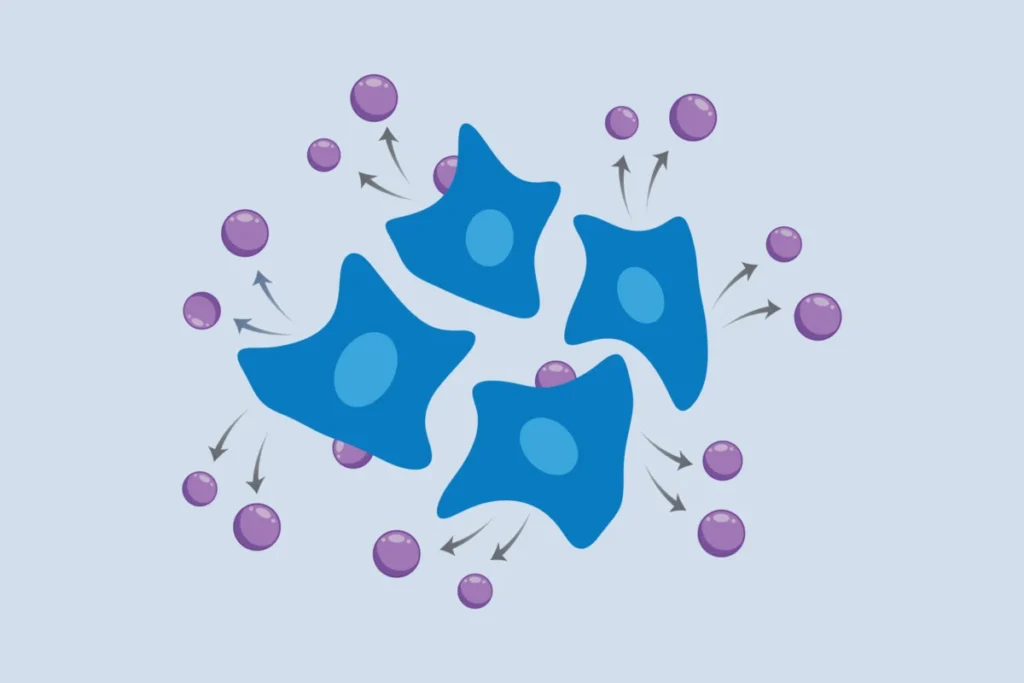
The breakthrough centers on the ingenious repurposing of virus-like particles (VLPs) as cellular messengers. Engineered cells are now capable of packaging their own RNA into these VLPs and releasing them into their surrounding culture medium. Scientists can then simply collect and analyze the RNA from the medium, enabling repeated, non-invasive measurements of gene activity from the same cell population over extended periods. This capability is poised to unlock deeper insights into how cells mature, respond to external stimuli, or aberrantly contribute to disease progression, as well as how they react to therapeutic interventions. The Broad Institute’s announcement on September 1 heralds a new era in longitudinal biological research, promising to shed light on the intricate, time-dependent orchestrations within living systems.
The Static Limitations of Traditional Transcriptomics
For decades, scientists have recognized the immense value of understanding a cell’s transcriptome – the complete set of RNA molecules produced by a cell. This intricate collection of messenger RNAs, ribosomal RNAs, transfer RNAs, and various non-coding RNAs serves as a direct readout of a cell’s genetic activity, providing critical clues about its identity, function, and state. The transcriptome dictates which proteins are being made, in what quantities, and in response to which internal or external cues, making it a cornerstone of modern biology and medicine.
Early methods for transcriptome analysis, such as Northern blotting and later microarrays, offered a snapshot of gene expression for thousands of genes simultaneously. The advent of RNA sequencing (RNA-seq) revolutionized the field, providing an unbiased, high-resolution quantitative measure of nearly all RNA transcripts present in a sample. More recently, single-cell RNA sequencing (scRNA-seq) has pushed the boundaries further, allowing researchers to profile gene expression at the resolution of individual cells, revealing previously hidden cellular heterogeneity within tissues and populations.
However, a pervasive and fundamental limitation has plagued all these traditional approaches: they require the lysis, or destruction, of the cell to extract its RNA content. This means that every measurement is a "snapshot" in time. To study dynamic processes – how a cell differentiates, responds to a drug, or progresses through a disease state – researchers have been forced to analyze different populations of cells sacrificed at various time points. This inferential approach, while powerful, inherently sacrifices the ability to track changes within the same individual cell or precisely the same population over time. It’s akin to trying to understand the narrative of a complex movie by only looking at a series of unrelated photographs taken from different scenes, rather than watching the film unfold. This inability to conduct longitudinal, non-destructive monitoring has created a significant gap in understanding the true kinetics and causal relationships governing cellular biology, particularly in processes that evolve over hours, days, or even weeks.
A Decade-Long Vision: From "Science Fiction" to Scientific Reality
The journey to overcome this fundamental hurdle began more than a decade ago in the laboratory of Paul Blainey, a core member of the Broad Institute and a professor of biological engineering at MIT. Blainey and his team were driven by the ambitious goal of developing a method for RNA sequencing that would not require the destruction of cells. "The existing methods were a bit medieval and involved stabbing cells or cutting pieces off of them," Blainey recalled, highlighting the inherent violence of traditional approaches.
At the time, the concept of a cell actively reporting its internal gene expression without intervention seemed almost fantastical. Blainey characterized their initial vision as "complete science fiction," underscoring the high-risk, high-reward nature of the endeavor. Yet, inspired by the rapid adoption and transformative impact of molecular technologies like CRISPR-based gene editing, Blainey and study first author Jacob Borrajo committed to pursuing a purely molecular solution. They recognized that while challenging and time-consuming, a molecularly encoded approach would ultimately be scalable and easily adaptable by other research laboratories, ensuring its widespread utility and impact.
The key to their innovative design lay in the natural world: retroviruses. These viruses have, over millions of years, evolved an incredibly efficient mechanism for packaging their RNA genomes into protective protein shells – virions – and releasing them from an infected cell to spread to others. This natural "delivery system" provided a compelling blueprint. The Broad and MIT team hypothesized that if they could engineer mammalian cells to express specific retroviral structural proteins, these proteins might be repurposed to encapsulate the cell’s own RNA, rather than viral RNA, and release it in a similar fashion. This idea represented a profound conceptual leap: transforming a mechanism of viral pathogenesis into a tool for biological discovery.
Unpacking the "Cellular Special Delivery": How it Works
The implementation of the "cellular self-reporting" method is a testament to sophisticated genetic engineering. The researchers engineered mammalian cells to express a specific retroviral structural protein. This protein is designed to integrate into the cell’s membrane, acting as a molecular scaffold. Crucially, it possesses the ability to recruit and bind to cellular RNA molecules, encapsulating them within a newly formed protein shell. This process creates what are known as virus-like particles (VLPs) – structures that mimic the exterior of a virus but contain only cellular RNA, lacking any viral genetic material or infectivity.
Once formed, these VLPs bud off from the cell membrane and are released into the liquid culture medium in which the cells are growing. The beauty of this system lies in its simplicity and non-invasiveness. Scientists can then simply collect a sample of the culture medium, isolate the VLPs, and extract the RNA contained within them. This RNA can then be sequenced using standard RNA-seq protocols, providing a comprehensive view of the transcriptome from that specific cell population. Critically, because the cells themselves remain alive and undisturbed throughout this process, researchers can perform this sampling repeatedly over time, generating a dynamic, longitudinal dataset of gene expression changes.
Co-first author Mohamad Najia, a research fellow in the Blainey lab and the lab of George Daley at Boston Children’s Hospital, highlighted the pragmatic advantages of this molecular approach. "Compared to methods using robotics or mechanical biopsies of cells, our molecularly encoded solution could be much more broadly enabling for the average life science or biomedical lab, particularly the time dynamic questions that we hope to elucidate with this technology," Najia stated. This ease of adoption is a crucial factor for the widespread integration of new technologies into the scientific community, distinguishing it from approaches that require specialized, expensive equipment or highly complex manual manipulations. Najia and Borrajo led the development alongside co-first author Anna Le, a postdoc in the Blainey lab.
Versatile Applications: A Broad Spectrum of Biological Systems
To demonstrate the broad applicability and robustness of their new method, the research team meticulously tested "cellular self-reporting" across a diverse array of cellular model systems. Their findings underscore its potential utility across various biological disciplines.
The method proved effective in:
- Immortalized human cells: Standard laboratory cell lines often used for fundamental research.
- Cancer cell lines: Essential for studying tumor biology, drug resistance, and therapeutic development.
- Stem cells and neuronal cells derived from them: Crucial for developmental biology, regenerative medicine, and neurological disease modeling.
- Primary cells from human donors: Offering more physiologically relevant insights for disease research and personalized medicine.
Beyond individual cell types, the team successfully applied the method to more complex co-culture systems. In experiments involving two different human cell types growing together, they incorporated specific tags onto the virus-like particles. These tags allowed scientists to distinguish and separately analyze the RNA signals originating from each cell type during sequencing, providing a nuanced understanding of intercellular communication and interaction dynamics within mixed populations. This capability is vital for studying tissues where multiple cell types interact to perform specific functions.
A particularly compelling demonstration of the method’s power involved its application to three-dimensional (3D) cellular structures, which are increasingly used to mimic in vivo tissue architecture and function more accurately than traditional 2D cultures. The researchers utilized spheroids of human endothelial cells – self-assembling clusters that better replicate the microenvironment of blood vessels. By applying "cellular self-reporting," they were able to capture short-term transcriptional changes in these spheroids after biochemical stimulation, demonstrating the method’s utility in systems where mechanical disruption would be highly detrimental to structural integrity and biological relevance.
Further expanding its scope, the Broad team collaborated with Linda Griffith, a professor of biological and mechanical engineering at MIT, to integrate the method with her lab’s cutting-edge organ-on-a-chip devices. These microfluidic systems are engineered to replicate the physiology and microenvironment of human organs, offering powerful platforms for disease modeling and drug screening with reduced reliance on animal models. However, the intricate design and small scale of these devices make retrieving cells for analysis extremely challenging. "Cellular self-reporting" provided an elegant solution. The researchers continuously monitored gene expression dynamics in endothelial cells within these organ-on-a-chip devices over time. Their experiments revealed significant changes in genes related to how tissues form vascular networks, specifically demonstrating that these dynamics were dependent upon the source of supporting fibroblasts – whether from the uterus or the lung. This finding not only validates the method’s efficacy in highly complex, physiologically relevant models but also highlights its capacity to uncover subtle, yet critical, biological differences influenced by tissue microenvironment.
Statements and Broader Scientific Anticipation
Paul Blainey articulated the driving philosophy behind his lab’s work: "Our lab focuses our time and resources on developing tools that will actually get used and make real impact on the broader field." He expressed profound satisfaction at the realization of this decade-long endeavor, emphasizing the journey from a seemingly impossible concept to a tangible scientific tool. "It’s so gratifying to see a real coming to fruition of this concept, which was complete science fiction when we started. It’s a great example of the innovative impact long-term high-risk, high-reward research can have." This sentiment resonates deeply within the scientific community, where the pursuit of audacious, transformative ideas often requires sustained effort and belief in the face of significant challenges.
The scientific community is likely to greet this development with considerable enthusiasm. Researchers grappling with the limitations of destructive assays in fields ranging from developmental biology to neurodegenerative disease research will recognize the immediate utility of a non-invasive, longitudinal transcriptomic platform. Pharmaceutical and biotechnology companies, in particular, may see immense potential for accelerating drug discovery and development. The ability to monitor cellular responses to drug candidates in real-time within complex in vitro models, such as organ-on-a-chip systems, could significantly enhance preclinical testing, providing more accurate predictors of efficacy and toxicity and potentially reducing the time and cost associated with bringing new therapies to market.
Unlocking the Dynamics of Life: Implications and Future Directions
The introduction of "cellular self-reporting" represents more than just a new technique; it heralds a paradigm shift in how dynamic biological processes can be investigated. Its implications span multiple critical areas of research and medicine:
- Disease Mechanisms: For diseases characterized by gradual progression or acute flares, such as cancer metastasis, neurodegenerative disorders like Alzheimer’s or Parkinson’s, autoimmune diseases, and chronic infections, the method offers an unprecedented window into the real-time molecular changes occurring within affected cells. Researchers can now track how cells "go awry over time," identifying critical transition points, early biomarkers of disease onset, or mechanisms of resistance to therapy in situ.
- Drug Discovery and Development: The ability to continuously monitor cellular responses to drug candidates in a non-destructive manner promises to revolutionize preclinical drug screening. It allows for the detailed study of dose-response kinetics, the identification of off-target effects, and the assessment of drug resistance mechanisms as they emerge over time. This could lead to the development of more effective and safer therapeutics, optimized for specific cellular contexts and patient populations. Furthermore, by providing more sophisticated in vitro models, it could help reduce the ethical and practical reliance on animal testing.
- Developmental Biology and Regenerative Medicine: Understanding how cells differentiate, form tissues, and regenerate damaged organs requires observing complex genetic programs unfolding over time. "Cellular self-reporting" provides the means to track lineage decisions, maturation processes, and the response of stem cells to differentiation cues without perturbing the developing system. This is invaluable for engineering tissues and organs for regenerative medicine applications.
- Personalized Medicine: In the future, this technology could facilitate the testing of individual patient-derived cells (e.g., in organoid models) against various drug regimens, allowing for real-time monitoring of their unique transcriptomic responses. This could pave the way for highly personalized treatment strategies, moving beyond a one-size-fits-all approach.
- Fundamental Biological Research: Beyond immediate translational applications, the method will empower basic scientists to ask and answer fundamental questions about cellular plasticity, stress responses, metabolic shifts, and environmental adaptations with an unprecedented level of temporal resolution. It transforms the study of cellular life from a series of static photographs into a continuous, unfolding movie.
The Broad team is actively pursuing new applications and biological questions that can be addressed with their system. A significant future direction is to adapt the approach for studying single cells, which would combine the temporal resolution of "cellular self-reporting" with the high spatial and cellular resolution of single-cell transcriptomics. For now, they extend an invitation to scientists across the globe who are interested in tracking how cells and tissues change over time to explore and adopt this transformative method. The journey from "science fiction" to a widely accessible and impactful research tool underscores the power of sustained, innovative research in pushing the boundaries of what is possible in biological discovery.