Researchers have made a significant breakthrough in the field of tissue engineering, demonstrating that the simple mechanical action of repeatedly jostling an artery can precisely stimulate the growth of new, smaller capillaries. This novel approach, detailed in a recent publication in the Proceedings of the National Academy of Sciences, reveals that the direction and degree of mechanical stretching can directly influence the growth patterns and density of these new blood vessels. This discovery offers scientists an unprecedented method to engineer artificial blood vessels and program their intricate growth patterns, addressing a long-standing challenge in regenerative medicine.
The intricate networks of blood vessels, particularly the microscopic capillaries, are the lifeblood of healthy tissues, responsible for delivering oxygen and nutrients while removing waste products. The ability to create and control such networks within engineered tissues has been a holy grail for scientists aiming to develop artificial organs, repair damaged tissues, and treat various diseases characterized by insufficient blood supply. Until now, state-of-the-art protocols have struggled to fabricate these highly organized vascular systems with the necessary precision and reproducibility.
Overcoming Hurdles in Vascularization
The challenge of vascularizing engineered tissues is multifaceted. While larger blood vessels like arteries and veins can be replicated using advanced 3D printing technologies, the creation of fine, thread-like capillary networks, essential for microcirculation, remains beyond the current capabilities of these methods. Traditional biological approaches often involve cultivating individual endothelial cells (the cells that line blood vessels) in nutrient-rich media, sometimes supplemented with chemical growth factors. However, controlling the precise architecture, density, and directionality of vessel growth using chemical cues alone has proven exceedingly difficult.
"Healthy tissues depend on organized blood vessel networks, but state-of-the-art protocols don’t enable fabricating such networks within engineered tissues," explains Ritu Raman, an associate professor of mechanical engineering at MIT and the lead author of the study. "You can try to pattern chemical cues, like growth factors, to direct where vessels grow, but you can’t do this very precisely. We thus need other types of patternable cues that can help us build tissues with organized vessels."
The research team, including MIT co-authors Sina Kheiri, Jessica Shah, Shashaank Venkatesh, and Roger Kamm, along with Peiyuan Chai and Ryan Flynn at Harvard University, posited that mechanical forces, rather than purely chemical signals, might offer a more precise and programmable means to guide vascular growth. This hypothesis stemmed from their previous successes in using mechanical stimulation to guide the growth of artificial muscles and nerves, suggesting a broader principle of mechanobiology at play.
A Novel "Blood-Vessel-on-a-Chip" Platform
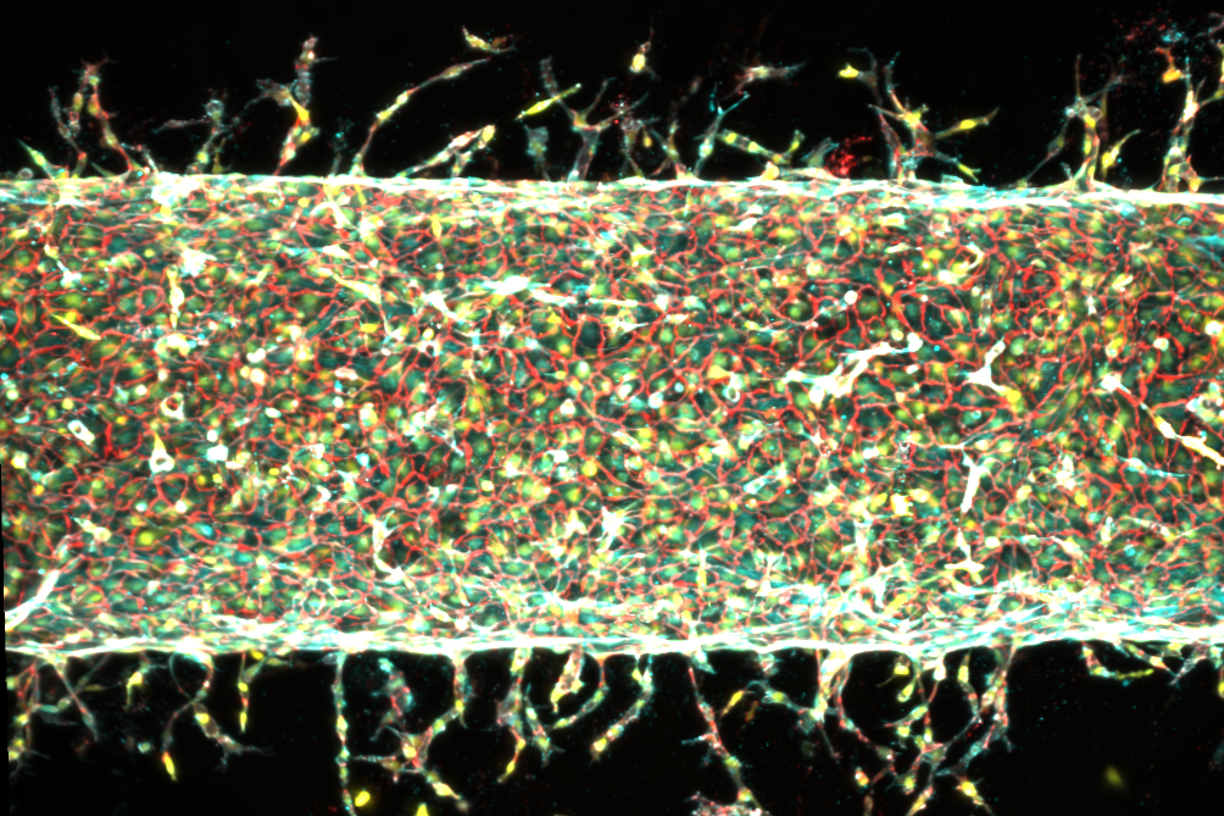
Building on their earlier work, Raman and her students developed a specialized microfluidic device, aptly named a "blood-vessel-on-a-chip." This innovative platform, smaller than a postage stamp, consists of a nutrient-rich gel embedded with a small magnet. A thin channel was created lengthwise through the gel and subsequently coated with live endothelial cells. These cells, given their natural propensity to form blood vessels, spontaneously began to sprout new, capillary-like structures into the surrounding gel, mimicking the early stages of angiogenesis (the formation of new blood vessels from pre-existing ones).
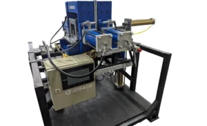
The ingenuity of the system lay in its mechanical control. The device was placed under a motorized stage fitted with small, suspended magnets. By precisely moving these external magnets, the researchers could manipulate the embedded magnet, thereby applying controlled mechanical forces—specifically, stretching and jostling—to the central artery within the gel. This setup allowed for meticulous observation of how the emerging blood vessels responded to varying directions and degrees of mechanical stimulation.
The Power of Mechanical Exercise: "Moving is Good"
The results were compelling. When the central artery was left undisturbed in the gel, it did sprout some new vessels, but their growth was largely random and unorganized. However, when the artery was subjected to mechanical jostling, a significant increase in the number of sprouting capillaries was observed.
The researchers systematically explored the effects of different stretching parameters:
- Increased Sprouting: When the gel was stretched back and forth by 5 percent of its total width, a substantial number of new vessels proliferated from the main artery.
- Controlled Length: Stretching the gel by a greater degree, specifically 15 percent, resulted in fewer vessels sprouting, but those that did emerge grew noticeably longer. This suggests a potential mechanism for controlling vessel length, a critical parameter in functional vascular networks.
- Directional Guidance: Crucially, when the team altered the direction of the mechanical stretching, the new vessels responded in kind, reorienting their growth to follow the imposed mechanical pattern. This demonstrated an unprecedented level of directional control over angiogenesis, a capability previously difficult to achieve with chemical methods alone.
"The main takeaway is: Stretching the blood vessel back and forth seems to enhance the number of new capillaries that grow," Raman noted. "We’re finding that moving is good, which is always the takeaway of everything we do in our lab. Mechanical forces play an important role in our bodies. That means that if you want to grow more or less vessels, or shorter or longer vessels, or vessels in certain directions, we now know how to do that."
This finding resonates with the broader understanding of mechanotransduction, the process by which cells sense and respond to mechanical stimuli. From bone remodeling in response to weight-bearing exercise to the development of the heart and blood vessels during embryogenesis, mechanical forces are fundamental regulators of cellular behavior and tissue development. This research provides a tangible demonstration of how these forces can be harnessed for bioengineering purposes.
Unveiling the Molecular Mechanism: The Role of PIEZO1
To delve deeper into the underlying biological mechanisms, the research team investigated the role of a specific gene: PIEZO1. This investigation was spurred by a serendipitous interaction between Ritu Raman and molecular biologist Ardem Patapoutian, who was awarded the Nobel Prize in Physiology or Medicine in 2021 for his groundbreaking discovery of ion channels that respond to mechanical pressure.
Patapoutian’s work identified PIEZO1 and PIEZO2 as key mechanosensitive ion channels embedded in cell membranes. These channels act as cellular gatekeepers, opening and closing in response to mechanical deformation, thereby regulating the flow of ions (such as calcium) into and out of the cell. This influx of ions triggers a cascade of intracellular signaling pathways that can influence various cellular processes, including growth, differentiation, and migration.
After Raman presented her group’s experimental results showing a clear link between blood vessel growth and mechanical stimulation, Patapoutian proposed that the PIEZO1 channel could be the missing link. He hypothesized that the mechanical exercise applied to the central artery might be stimulating these ion channels in the endothelial cells to open, thereby initiating the signaling cascade that promotes new blood vessel growth.
To test this hypothesis, the team employed gene-editing techniques to suppress the activity of the PIEZO1 gene in endothelial cells. If PIEZO1 was indeed the primary mediator of mechanical stimulation, then reducing its activity should inhibit the observed vessel growth. The results unequivocally supported Patapoutian’s suggestion. When experiments were repeated with endothelial cells genetically modified to have reduced PIEZO1 expression, significantly fewer new blood vessels sprouted, even under mechanical exercise. This critical finding firmly established PIEZO1 as a crucial gatekeeper in mechanically induced angiogenesis.
Broader Implications and Future Directions
The ability to precisely program blood vessel growth using physical cues represents a paradigm shift in tissue engineering and regenerative medicine. The implications of this research are vast and transformative:
- Artificial Organ Development: One of the most significant hurdles in creating functional artificial organs (e.g., kidneys, livers, hearts) for transplantation is the lack of an integrated vascular supply. This discovery offers a scalable and reproducible method to build organized vessel networks within engineered tissues, bringing the dream of functional artificial organs closer to reality. Such vascularized constructs could potentially bypass the need for organ donors and address the critical shortage of transplantable organs.
- Treating Ischemic Diseases: Diseases like heart attack, stroke, and peripheral artery disease are characterized by insufficient blood flow to tissues due to blocked or damaged vessels. This technology could lead to novel therapeutic strategies to stimulate new vessel growth (therapeutic angiogenesis) in affected areas, restoring blood supply and improving tissue function.
- Enhanced Wound Healing: Chronic wounds, such as diabetic ulcers and severe burns, often struggle to heal due to inadequate blood supply. The ability to precisely direct the growth of new capillaries could significantly accelerate wound healing, improve tissue regeneration, and reduce complications.
- Disease Modeling and Drug Discovery: Creating more physiologically relevant in vitro models of human tissues, complete with functional vascular networks, would revolutionize drug testing and disease modeling. Researchers could study disease progression and test new therapies in environments that more closely mimic the human body, leading to more accurate predictions and efficient drug development.
- Understanding Cancer Biology: The uncontrolled growth of blood vessels (angiogenesis) is a hallmark of cancer, supplying tumors with nutrients. Understanding and potentially manipulating mechanical cues could offer new avenues for anti-angiogenic therapies to starve tumors.
The research team is already looking ahead to the next steps. "We are now investigating how precisely patterning blood vessel growth can help improve muscle function," says co-author Jessica Shah, highlighting the interdisciplinary nature of their work and the potential to integrate vascular networks with other engineered tissues like muscles and nerves.
This work was supported, in part, by the U.S. Department of War Army Research Office Early Career Program and PECASE Grant, and a Department of War DURIP Program Grant, underscoring the strategic importance of this research for both civilian and military applications, particularly in trauma care and rehabilitation. The integration of mechanobiology with tissue engineering heralds a new era where mechanical forces, long appreciated for their role in biological development, can be precisely orchestrated to build and repair the human body, offering profound hope for patients suffering from a wide array of debilitating diseases and injuries.