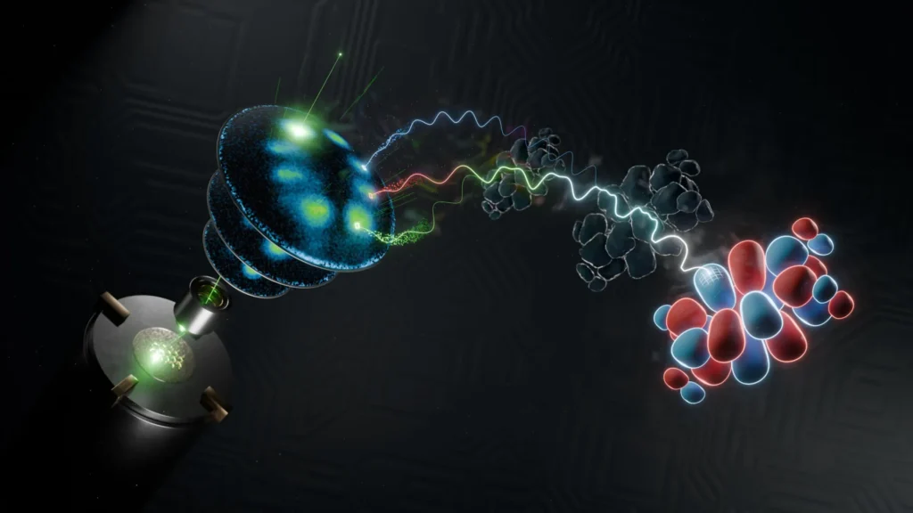
The field of quantum mechanics has reached a significant milestone as an interdisciplinary research team at the University of Göttingen successfully captured a complete three-dimensional image of a molecular wavefunction. This achievement, recently detailed in the journal Nature Communications, marks a transition from treating the wavefunction as a purely mathematical abstraction to utilizing it as a visible, measurable tool for understanding the internal dynamics of organic molecules. By merging high-resolution photoelectron spectroscopy with a revolutionary reconstruction algorithm, the researchers have bypassed long-standing technical barriers that previously restricted such observations to massive, multi-billion-dollar synchrotron facilities.
For over a century, the wavefunction has served as the cornerstone of quantum theory. Unlike classical objects that possess a definite position and velocity, fundamental particles like electrons exist in a state of probability. The wavefunction, denoted by the Greek letter psi (Ψ), provides the mathematical framework to calculate these probabilities. Within the complex environment of a molecule, these wavefunctions manifest as "molecular orbitals"—the specific spatial distributions that electrons occupy. These orbitals are the "blueprints" of chemical behavior; they dictate how a molecule bonds with others, how it reacts to light, and how it conducts electricity. Despite their fundamental importance, wavefunctions are not directly observable through conventional microscopy because the act of measurement typically collapses the quantum state or fails to capture the essential "phase" information required for a full 3D reconstruction.
The Quantum Challenge: Capturing the Unobservable
The primary difficulty in imaging a wavefunction lies in its dual nature. A complete description of an electron’s state requires both its amplitude (which relates to the probability of finding the electron in a certain spot) and its phase (which describes the wave-like oscillation of the particle). While experimental techniques have long been able to measure the intensity or amplitude of electron distributions, the phase information is often lost during detection—a dilemma known in physics as the "phase problem."
To overcome this, the Göttingen team employed an indirect measurement strategy known as photoemission orbital tomography (POT). In this process, the molecule is hit with high-energy photons, causing it to eject electrons. By measuring the momentum of these emitted photoelectrons, scientists can gain a "momentum-space" map of the molecular orbital. However, this map is essentially a 2D projection and represents only half of the necessary information. To transform this data into a 3D image of the wavefunction in "real space," the researchers had to solve the missing phase information and account for the third spatial dimension, a task that has historically required exhausting amounts of data and access to massive particle accelerators.
Professor Stefan Mathias, a lead researcher at the University of Göttingen, emphasized the philosophical and practical weight of this hurdle. "The wavefunction is a fundamental quantity in quantum mechanics, yet it cannot be directly observed or measured," Mathias noted. He explained that the team’s success relied on a hybrid approach: using physics to gather the raw data and sophisticated mathematics to fill in the gaps that nature hides from direct view.
A Chronology of Molecular Imaging Progress
The journey toward 3D wavefunction imaging has been decades in the making. In the early 20th century, X-ray crystallography allowed scientists to deduce the positions of atoms within a crystal lattice, but it could not visualize the electron clouds (orbitals) themselves. By the 1960s and 70s, the development of photoelectron spectroscopy provided the first glimpses into electron energy levels, yet spatial imaging remained elusive.
The early 2000s saw the birth of orbital tomography, where researchers used synchrotrons—circular particle accelerators miles in circumference—to produce the intense X-ray beams needed to knock electrons out of molecules with enough precision to map their orbitals. While successful, these experiments were limited. Synchrotron time is highly competitive and expensive, and the sheer volume of data required to move from 2D slices to a 3D volume meant that imaging a single molecule could take days or weeks of continuous measurement.
The breakthrough at Göttingen represents the latest and most significant entry in this timeline. By developing a new class of algorithms and utilizing a compact, laboratory-based light source, the team has compressed a process that once required a football-stadium-sized facility into a setup that fits within a standard university laboratory. This shift not only democratizes the technology but also opens the door to "time-resolved" imaging, where changes in the wavefunction can be tracked in real-time.
Technical Synergy: Soft X-Rays and Algorithmic Reconstruction
The success of the study rests on two primary technological pillars: a high-harmonic generation (HHG) light source and a ground-up redesign of reconstruction mathematics.
The light source used by the team is a lab-based soft X-ray generator. This device uses ultra-fast laser pulses to create "harmonics"—multiples of the laser’s frequency—resulting in extremely short bursts of soft X-ray radiation. These pulses are measured in femtoseconds (one quadrillionth of a second). The brevity of these pulses is critical; it allows the researchers to "freeze" the motion of electrons, providing a sharp snapshot of the molecular orbital before the system can shift or decohere.
Complementing this hardware is a new mathematical algorithm designed by Dr. Matthijs Jansen and his colleagues. Traditional reconstruction methods required the molecule to be rotated and measured at dozens of different angles to build a 3D picture, similar to how a medical CT scan works. The Göttingen team’s algorithm, however, is far more efficient. By utilizing "prior knowledge" about the physical constraints of molecular systems, the algorithm can reconstruct the 3D wavefunction from significantly less experimental data.
"We introduce two powerful new concepts," explained Dr. Jansen. "First, by redesigning the computer algorithm from the ground up, reliable 3D images can now be obtained using much less experimental data. Second, the experiment is based upon a powerful, lab-based soft-X-ray light source that provides ultrashort light pulses. It is the combination of these two techniques that has this remarkable impact."
Achieving Sub-Atomic Resolution
The precision of the resulting images is staggering. The team was able to distinguish features within the molecular orbital that are smaller than the distance between individual carbon atoms. In the world of organic chemistry, carbon-carbon bonds typically measure about 0.15 nanometers. The Göttingen imaging technique provides a resolution that penetrates deep into the "internal anatomy" of the molecule, showing how electron density pools and thins between atomic nuclei.
This level of detail is essential for understanding "functional" molecules, such as those used in organic light-emitting diodes (OLEDs) or organic solar cells. In these materials, the efficiency of light emission or energy conversion is determined by the exact shape of the molecular orbitals. Even a slight distortion in the wavefunction can significantly alter the material’s performance. By seeing the 3D structure clearly, scientists can now verify theoretical models with unprecedented accuracy.
The Future of "Molecular Movies"
While the 3D static imaging of a wavefunction is a landmark achievement, the researchers are already looking toward the next frontier: four-dimensional imaging, where the fourth dimension is time.
Dr. Wiebke Bennecke, the study’s first author, highlighted the potential for "stroboscopic videography" at the atomic scale. "This technique might mean that stroboscopic videography becomes a reality, allowing us to observe not just the shape of wavefunctions, but also to see how it changes with ultrafast resolution," Bennecke stated.
The goal is to capture "molecular movies" with femtosecond resolution. At this timescale, researchers could watch a chemical reaction happen frame-by-frame. They could observe how a molecule’s wavefunction deforms when it absorbs a photon, how it redistributes its charge during a chemical bond rupture, or how it interacts with an incoming catalyst. This would move chemistry from a science of "before and after" to a science of "during," providing a masterclass in how to control matter at its most fundamental level.
Broader Implications and Scientific Analysis
The implications of this research extend far beyond the laboratory. In the pharmaceutical industry, the ability to visualize the 3D wavefunctions of drug molecules as they interact with protein receptors could lead to more effective and targeted therapies. In the field of electronics, understanding the 3D orbitals of semi-conducting molecules is vital for the development of next-generation molecular computers and high-efficiency batteries.
Furthermore, the shift from synchrotrons to lab-based sources is a paradigm shift for quantum chemistry. It allows for rapid iteration. A chemist could potentially synthesize a new molecule and image its electronic structure in the same week, rather than waiting months for a scheduled slot at a national facility. This acceleration of the feedback loop between synthesis and characterization is likely to trigger a surge in materials discovery.
From a theoretical perspective, this work provides a rigorous test for quantum mechanical simulations. Computational chemists often use "Density Functional Theory" (DFT) to predict molecular behavior. While DFT is powerful, it involves approximations. The high-resolution 3D images produced at Göttingen provide a "ground truth" that can be used to refine these computational models, making future simulations more reliable.
Conclusion: A New Era of Quantum Visibility
The University of Göttingen’s achievement represents a rare moment where a theoretical concept—the 3D molecular wavefunction—becomes a tangible piece of data. By successfully integrating soft X-ray physics with advanced data science, the team has not only mapped the invisible but has also provided a blueprint for the future of ultrafast science.
As the team continues to refine their lab-based setup, the transition from static 3D images to dynamic 3D videos appears inevitable. The ability to observe and eventually control the femtosecond-scale adaptations of molecules to their environment will likely redefine our understanding of chemical reactions, potentially ushering in a new era of "quantum-controlled" technology where interactions involving only a few atoms can be steered with absolute precision. For now, the scientific community celebrates a major victory: the wavefunction is no longer just a ghost in the equations; it is an image on the screen.