Imagine a future where doctors can precisely deposit microscopic capsules, laden with regenerative cells, directly into the intricate network of a beating heart to initiate tissue repair. This groundbreaking vision is now a significant step closer to reality thanks to a pioneering method developed by a team of scientists at the California Institute of Technology (Caltech). Their innovative technique allows for the 3D printing of polymers at specific, deep-seated locations within living animals, leveraging the power of sound for unparalleled precision. This advancement has already demonstrated its efficacy in delivering targeted drug therapies and sealing internal wounds with a bioadhesive "glue."
A Leap Beyond Surface-Level Treatments
For years, medical researchers have explored methods to trigger the polymerization process – the crucial linking of monomer units to form larger polymer chains – within living organisms. Previous successful attempts often relied on infrared light. However, this approach suffered from a critical limitation: its shallow penetration depth. "Infrared penetration is very limited. It only reaches right below the skin," explained Wei Gao, a professor of medical engineering at Caltech and a Heritage Medical Research Institute Investigator. This constraint severely restricted the application of such techniques to superficial tissues.
The Caltech team’s breakthrough overcomes this significant hurdle. "Our new technique reaches the deep tissue and can print a variety of materials for a broad range of applications, all while maintaining excellent biocompatibility," stated Professor Gao. This ability to access and manipulate materials deep within the body opens up a vast new frontier for minimally invasive medical procedures.
The Science Behind the Sound: A Novel Hybrid Approach
The culmination of this research, detailed in a recent publication in the esteemed journal Science, describes the novel in vivo 3D-printing technique. Beyond its applications in delivering drugs and cells, the study also highlights the printing of bioelectric hydrogels. These advanced polymers are embedded with conductive materials, paving the way for internal monitoring of vital physiological signs, akin to electrocardiograms (ECGs).
The lead author of the study, Elham Davoodi, an assistant professor of mechanical engineering at the University of Utah, spearheaded the crucial work during her tenure as a postdoctoral scholar at Caltech. Her expertise was instrumental in combining established biomedical tools with an entirely new triggering mechanism.
The Genesis of an Innovative Concept
The quest for deep-tissue in vivo printing necessitated a robust localization mechanism. Recognizing the established prowess of ultrasound in penetrating deep into biological tissues, the Caltech team opted to build upon this well-understood platform. However, the primary challenge remained: how to precisely trigger the crosslinking of monomers at a specific point in time and space, and only when intentionally activated.
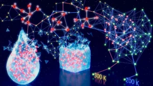
Their ingenious solution involved a sophisticated synergy between ultrasound and low-temperature-sensitive liposomes. Liposomes, essentially microscopic, spherical vesicles encased in protective fat layers, are already a familiar tool in drug delivery. In this novel application, the researchers loaded these liposomes with a crosslinking agent – the chemical key that initiates polymerization. These drug-laden liposomes were then suspended within a "bioink," a composite material comprising the monomers of the desired polymer, an imaging contrast agent to visualize the polymerization process, and, crucially, the therapeutic payload. This payload could be a pharmaceutical drug, but also, as the researchers demonstrated, therapeutic cells or conductive materials like carbon nanotubes or silver, enabling a wide array of functional materials. This carefully formulated bioink was then injected directly into the animal’s body.
A Subtle Temperature Shift to Ignite Printing
The defining characteristic of the loaded liposomes is their sensitivity to temperature. By focusing ultrasound waves onto a targeted region, the scientists can induce a localized temperature increase of approximately 5 degrees Celsius. This seemingly small change is sufficient to destabilize the liposome membranes, triggering the release of their encapsulated crosslinking agent. "Increasing the temperature by a few degrees Celsius is enough for the liposome particle to release our crosslinking agents," Professor Gao elaborated. "Where the agents are released, that’s where localized polymerization or printing will happen." This highly localized release mechanism ensures that printing occurs exclusively at the intended site, minimizing collateral effects and maximizing therapeutic precision.
Visualizing the Unseen: Ultrasound-Guided Polymerization
To confirm and guide the printing process, the team incorporated a clever imaging strategy. They utilized gas vesicles derived from bacteria as an imaging contrast agent. These microscopic, air-filled protein capsules exhibit a strong signal on ultrasound imaging. More importantly, they are exquisitely sensitive to the chemical environment. As the liquid monomer solution undergoes crosslinking to form a solid polymer gel network, a chemical transformation occurs. This transformation directly affects the gas vesicles, causing a detectable change in their ultrasound contrast. This dynamic change allows scientists to precisely monitor, in real-time, when and where polymerization is occurring, enabling them to sculpt intricate patterns within the living animal.
The researchers have aptly named their groundbreaking technology the "Deep Tissue In Vivo Sound Printing" (DISP) platform.
Early Successes and Promising Clinical Trajectories
The potential of the DISP platform was underscored by an early experimental success. When the team employed the system to print doxorubicin-loaded hydrogels near a bladder tumor in mice, they observed a substantially greater rate of tumor cell death over several days compared to control groups that received the drug via conventional direct injection. This outcome suggests a significant improvement in drug delivery efficacy and targeted therapeutic action.
"We have already shown in a small animal that we can print drug-loaded hydrogels for tumor treatment," Professor Gao remarked. The team’s immediate future plans involve translating this success to larger animal models, with the ultimate aspiration of evaluating the DISP platform’s potential in human clinical trials in the not-too-distant future.
The Future of Precision Medicine: AI and Autonomous Printing
Looking further ahead, the Caltech team envisions an even more sophisticated integration of artificial intelligence (AI) with the DISP platform. Machine learning algorithms could significantly enhance the system’s ability to autonomously identify and target specific anatomical locations, further refining the precision of focused ultrasound application.
"In the future, with the help of AI, we would like to be able to autonomously trigger high-precision printing within a moving organ such as a beating heart," Professor Gao stated, painting a picture of advanced, automated medical interventions. This integration of AI promises to unlock the full potential of the DISP platform for complex surgical scenarios and the treatment of dynamic biological systems.
The research was generously supported by funding from the National Institutes of Health, the American Cancer Society, the Heritage Medical Research Institute, and the Challenge Initiative at UCLA. Crucial microscopy work was conducted at the Advanced Light Microscopy/Spectroscopy Laboratory and the Leica Center of Excellence at the California NanoSystems Institute at UCLA, highlighting the collaborative and resource-intensive nature of such pioneering scientific endeavors.
Broader Implications and Potential Impact
The development of the DISP platform represents a paradigm shift in the field of regenerative medicine and targeted therapeutics. The ability to precisely deliver therapeutic agents, cells, or even structural materials deep within the body opens up a multitude of possibilities for treating a wide spectrum of diseases and injuries that are currently difficult or impossible to address with existing technologies.
Regenerative Medicine: The precise delivery of stem cells or other regenerative agents directly to damaged tissues, such as cardiac muscle after a heart attack or cartilage in an osteoarthritic joint, could revolutionize healing processes. By creating an optimal microenvironment and providing the building blocks for repair exactly where they are needed, the DISP platform could significantly enhance the body’s natural regenerative capabilities.
Targeted Cancer Therapies: The successful demonstration of enhanced tumor cell death with doxorubicin suggests a powerful new avenue for cancer treatment. By concentrating chemotherapy drugs directly at tumor sites, the DISP platform could reduce systemic toxicity, thereby improving patient quality of life and potentially increasing treatment efficacy. This precision delivery also holds promise for localized gene therapy or immunotherapy applications within tumors.
Wound Closure and Tissue Engineering: The reported use of bioadhesive polymers for sealing internal wounds highlights another critical application. This could lead to less invasive surgical procedures, faster recovery times, and reduced risks of infection and leakage. Furthermore, the ability to print complex polymer structures in situ could form scaffolds for tissue regeneration, guiding the growth of new tissue with unparalleled architectural control.
Biomedical Monitoring: The development of bioelectric hydrogels for physiological monitoring is a significant step towards implantable, long-term biosensors. These could provide continuous, real-time data on vital signs, offering early detection of disease exacerbations or critical physiological changes, particularly in patients with chronic conditions.
Challenges and Future Directions:
While the DISP platform shows immense promise, several challenges lie ahead. Scaling the technology for human use will require rigorous testing for safety, efficacy, and long-term biocompatibility. Developing a comprehensive library of printable materials with diverse functionalities will be crucial for a wide range of clinical applications. Furthermore, refining the AI integration to ensure absolute safety and reliability in complex, dynamic biological environments will be paramount.
The journey from laboratory discovery to widespread clinical adoption is often a long and arduous one. However, the fundamental breakthrough achieved by the Caltech team represents a critical leap forward. The DISP platform, by marrying the precision of sound-based localization with the versatility of 3D printing, has laid the foundation for a new era of minimally invasive, highly targeted medical interventions, promising to reshape the landscape of healthcare and patient outcomes for generations to come.