A groundbreaking discovery by researchers at the Massachusetts Institute of Technology (MIT) has upended long-held beliefs in laser physics, revealing a novel method for generating exceptionally precise and stable laser beams through self-organization within multimode optical fibers. This unexpected phenomenon, where increased laser power leads to a highly organized, needle-sharp beam rather than chaotic scattering, promises to revolutionize biomedical imaging, particularly for challenging structures like the human blood-brain barrier. The findings, detailed in a paper published today in Nature Methods, introduce a simpler yet more powerful approach to optical microscopy, offering unprecedented speed and clarity for observing biological processes in real-time.
A Paradigm Shift in Laser Physics
For decades, the prevailing understanding within the optics community dictated that intensifying the power within certain types of lasers, especially those transmitted through multimode optical fibers, would inevitably result in a chaotic and disordered light beam. This disarray stems from the inherent imperfections and multiple pathways light can take within such fibers, leading to scattering and a loss of focus. Researchers traditionally relied on complex and often expensive external beam-shaping components and intricate light engineering techniques to counteract this disorder, particularly when aiming for high-power, focused beams essential for advanced applications.
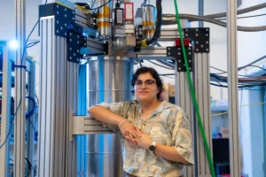
However, a team led by Sixian You, an assistant professor in the MIT Department of Electrical Engineering and Computer Science (EECS) and a member of the Research Laboratory for Electronics, has demonstrated that this conventional wisdom is not absolute. "The common belief in the field is that if you crank up the power in this type of laser, the light will inevitably become chaotic. But we proved that this is not the case," You stated, underscoring the counterintuitive nature of their discovery. "We followed the evidence, embraced the uncertainty, and found a way to let the light organize itself into a novel solution for bioimaging." This self-organizing principle represents a significant departure from traditional active beam manipulation, offering a pathway to high-quality beams with simplified setups.
The research paper’s lead author is Honghao Cao, an EECS graduate student. He is joined by fellow EECS graduate students Li-Yu Yu and Kunzan Liu; postdocs Sarah Spitz, Francesca Michela Pramotton, and Federico Presutti; Zhengyu Zhang PhD ’24; Subhash Kulkarni, an assistant professor at Harvard University and the Beth Israel Deaconess Medical Center; and Roger Kamm, the Cecil and Ida Green Distinguished Professor of Biological and Mechanical Engineering at MIT. Their collaborative effort spans electrical engineering, computer science, and biological and mechanical engineering, highlighting the interdisciplinary nature of modern scientific breakthroughs.
The Genesis of an Unexpected Discovery
The journey to this remarkable finding began with an observation that initially perplexed the research team. The MIT group had previously developed a sophisticated "fiber shaper," a device designed to precisely tune laser light passing through a multimode optical fiber. Multimode fibers, characterized by their wider core diameter compared to single-mode fibers, are capable of carrying a significantly higher amount of optical power. This makes them attractive for various applications, but their inherent tendency to support multiple light paths, or modes, makes it challenging to maintain a focused, coherent beam, especially at high power. The light often scatters, leading to a blurry or speckled output.
Graduate student Honghao Cao was systematically testing the limits of these multimode fibers, pushing them to extreme power levels to understand their endurance. His objective was to determine the maximum power a fiber could withstand before succumbing to damage or producing an unmanageably disordered beam. In typical scenarios, as power increases, the light experiences greater nonlinear interactions with the fiber material, exacerbating imperfections and leading to a more scattered and diffuse beam. This scattering is a well-documented phenomenon that limits the utility of high-power lasers in precise applications.
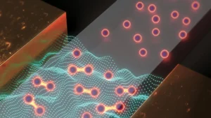
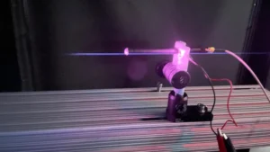
However, as Cao incrementally increased the laser power, approaching the very threshold where the fiber might burn out, he witnessed something entirely unexpected. Instead of becoming more disordered and scattered, the light beam underwent a dramatic transformation: it spontaneously collapsed into a single, intensely focused, needle-sharp beam. This observation defied all conventional expectations and sparked intensive investigation. "Disorder is intrinsic to these fibers. The light engineering you typically need to do to overcome that disorder, especially at high power, is a longstanding hassle," You explained. "But with this self-organization, you can get a stable, ultrafast pencil beam without the need for custom beam-shaping components." The implications of achieving such a precise beam without complex external optics were immediately apparent.
Unlocking the Secret: Two Critical Conditions
To understand and replicate this surprising phenomenon, the researchers meticulously identified two crucial, yet relatively simple, conditions that must be met. These conditions, when combined, create a unique optical environment where the light’s intrinsic properties enable self-organization.
First, the laser light must enter the multimode fiber at a precise, perfect zero-degree angle relative to the fiber’s central axis. This requirement is significantly more rigorous than typical alignment procedures for multimode fibers, which often tolerate slight angular deviations due to their larger core and power-carrying capacity. Conventional wisdom suggests that such precise alignment is less critical for multimode fibers compared to single-mode fibers.
Second, and perhaps most critically, the laser power must be progressively dialed up until the light begins to strongly interact with the glass material of the fiber itself. This interaction triggers what physicists refer to as "nonlinear optical effects." At lower power levels, light propagates through glass largely linearly, meaning its properties don’t significantly alter the medium or vice-versa. However, at sufficiently high intensities, the refractive index of the glass—its ability to bend light—changes in response to the light’s presence. This nonlinear response becomes a dominant factor.
"At this critical power, the nonlinearity can counter the intrinsic disorder, creating a balance that transforms the input beam into a self-organized pencil beam," Cao elaborated. This balance is the key. The nonlinear effects, rather than contributing to chaos, actively work to counteract the scattering caused by fiber imperfections. In essence, the light itself reshapes its propagation path, organizing into a stable, tightly focused beam. It’s a testament to the complex interplay between light intensity and material properties that can emerge under specific conditions.
The reason this phenomenon remained undiscovered for so long lies in the conventional experimental practices. Researchers typically operate at much lower power levels in multimode fibers to avoid the risk of damaging the fiber, or because the assumed chaotic behavior at higher powers made further investigation seem unproductive. Furthermore, the extreme precision in on-axis alignment was not deemed necessary for general multimode fiber applications. It was the combination of pushing power limits and meticulous alignment that unlocked this hidden behavior. The beauty of this method, as You points out, is its accessibility: "That is the charm of this method — you could do this with a normal, optical setup and without much domain expertise." This simplicity could significantly lower the barrier to entry for advanced optical imaging.
A Superior Beam for Precision Imaging
Once the researchers had successfully replicated and understood the conditions for generating this self-organized pencil beam, they proceeded to characterize its properties. The results were striking. The newly formed beam exhibited remarkable stability and a significantly higher resolution compared to many conventional beams used in optical microscopy. A common issue with traditional laser beams, particularly when attempting to achieve high power and focus, is the presence of "sidelobes"—blurry halos of light surrounding the main beam that can severely distort images and reduce contrast. The MIT team’s pencil beam, however, was notably pristine and tightly focused, largely free of these detrimental sidelobes. This characteristic makes it ideal for applications demanding exceptional clarity and precision.
Revolutionizing Bioimaging: The Blood-Brain Barrier
Building upon the robust characterization of their novel pencil beam, the MIT researchers wasted no time in demonstrating its practical utility in a critically important biomedical application: imaging the human blood-brain barrier (BBB). The BBB is a highly selective, tightly packed layer of endothelial cells that lines the brain’s capillaries, forming a formidable protective shield against circulating toxins and pathogens. While essential for brain health, this barrier also poses a major challenge for medicine, blocking the entry of approximately 98% of small-molecule drugs and nearly 100% of large-molecule drugs, including many promising therapies for neurological disorders and brain cancers.
Understanding how drugs interact with and potentially traverse the blood-brain barrier is paramount for pharmaceutical research and neurological treatment. Scientists and clinicians urgently need methods to visualize the dynamics of drug flow within the vasculature of the BBB and determine whether therapeutic agents effectively reach their intended targets within the brain. Current optical imaging techniques face significant limitations in this domain. As You explained, "with standard optical settings, the best one can do is capture one 2D section of the vasculature at a time, and then repeat the process multiple times to generate a fuller image." This process is time-consuming, provides limited dynamic information, and often requires the use of fluorescent tags, which can alter cellular behavior or introduce phototoxicity.
The new self-organized pencil beam offers a powerful solution to these challenges. By leveraging its ultrafast, high-precision capabilities, the researchers were able to dynamically track how cells absorb proteins in real-time within an in vitro human blood-brain barrier model. This capability represents a significant leap forward. "The pharmaceutical industry is especially interested in using human-based models to screen for drugs that effectively cross the barrier, as animal models often fail to predict what happens in humans," noted Roger Kamm, a co-author on the paper and a leading expert in bioengineering.
Perhaps one of the most transformative aspects of this new method is its ability to operate without the need for fluorescent tags. This label-free imaging capability is a "game-changer," according to Kamm. "For the first time, we can now visualize the time-dependent entry of drugs into the brain and even identify the rate at which specific cell types internalize the drug." This means researchers can observe natural cellular processes without interference from external markers, providing a more accurate representation of biological reality.
The team successfully captured cellular-level 3D images of unprecedented quality, generating these images approximately 25 times faster than with conventional methods. This combination of speed and resolution is crucial for observing dynamic biological events. "Usually, you have a tradeoff between image resolution and depth of focus — you can only probe so far at a time," You elaborated. "But with our method, we can overcome this tradeoff by creating a pencil-beam with both high resolution and a large depth of focus." This ability to maintain high resolution across a greater depth is critical for imaging complex 3D biological structures without having to repeatedly adjust focus or stitch together multiple shallow images.
The utility of this innovative approach extends beyond the blood-brain barrier. Sarah Spitz, a postdoc involved in the research, affirmed, "Importantly, however, this approach is not limited to the blood-brain barrier but enables time-resolved tracking of diverse compounds and molecular targets across engineered tissue models, providing a powerful tool for biological engineering." This broad applicability suggests the technique could become a staple in various areas of biomedical research, from studying cell-drug interactions in other organs to analyzing tissue development and disease progression.
Broader Implications and Future Directions
The scientific community has already recognized the potential impact of this discovery. Frank Wise, the Samuel B. Eckert Professor of Engineering Emeritus at Cornell University, who was not involved in the MIT research, commented, "You’s group realized this beam that concentrates energy in time and space could be valuable for microscopy techniques that depend on the intensity of the light that illuminates the sample. They demonstrated just that and found advantages over ordinary laser beams for imaging. It will be scientifically interesting to fully understand the creation of the new pencil beams, which could find use in a variety of imaging applications." Wise’s statement highlights both the immediate practical utility and the deeper scientific questions this discovery raises.
Looking ahead, the MIT researchers plan to delve deeper into the fundamental physics underpinning the self-organization of the pencil beam. A more comprehensive theoretical understanding could lead to further optimization and new applications. They also intend to apply the technique to other challenging imaging scenarios, such as visualizing neuronal activity within the brain, where high resolution, speed, and minimal invasiveness are paramount. The long-term vision includes working toward commercializing the technology, making this powerful new imaging tool accessible to a broader scientific and clinical audience.
The development of this self-organizing laser beam represents a significant leap forward in optical science and its application to biology. By challenging established norms and embracing unexpected experimental outcomes, the MIT team has not only unveiled a new physical phenomenon but also provided a potent new instrument for exploring the intricate dynamics of life, promising to accelerate discoveries in drug development, neuroscience, and beyond.
This pioneering work received substantial support from various entities, including MIT startup funds, Novo Nordisk Research Development, a National Science Foundation (NSF) CAREER Award, CZI Dynamic Imaging from the Chan Zuckerberg donor-advised fund through the Silicon Valley Community Foundation, the Manton Foundation, and the Fairbairn Menstruation Science Fund, underscoring the collaborative effort required for such transformative scientific endeavors.