CAMBRIDGE, MA – September 1, 2026 – In a significant scientific advancement poised to revolutionize the study of cellular biology and disease, researchers at the Broad Institute of MIT and Harvard, in collaboration with MIT, have developed a groundbreaking "cellular self-reporting" method that allows scientists to monitor a cell’s genetic activity over time without destroying the cell. This innovative approach, detailed in a recent publication in the prestigious journal Cell, marks a paradigm shift from traditional destructive methods, enabling an unprecedented dynamic view of cellular processes.
For years, scientists have relied on transcriptomics – the measurement of all RNA molecules produced by a cell – to decipher cellular identity, function, and responses to various stimuli. The transcriptome acts as a comprehensive readout of a cell’s active genes, providing critical insights into its current state. However, existing methods, most notably single-cell RNA sequencing (scRNA-seq), necessitate the lysis, or destruction, of cells to access their internal RNA content. While immensely powerful for capturing snapshots of cellular states at specific points in time, these techniques inherently preclude the observation of dynamic changes within the same cell population over an extended period. The inability to track individual cells or populations through developmental stages, disease progression, or therapeutic interventions has long been a significant bottleneck in biomedical research.
The newly introduced "cellular self-reporting" technology directly addresses this fundamental limitation. Spearheaded by senior author Paul Blainey, a core member of the Broad Institute and a professor of biological engineering at MIT, along the lead efforts of co-first authors Jacob Borrajo, Mohamad Najia, and Anna Le, the method leverages engineered virus-like particles (VLPs). These VLPs are utilized by living cells to autonomously package and secrete their RNA into the surrounding culture medium. Scientists can then simply sample this medium repeatedly, isolate the secreted RNA, and sequence it to reconstruct the cellular transcriptome, all while the cells remain viable and undisturbed. This non-invasive, longitudinal monitoring capability promises to unlock a deeper understanding of how gene activity evolves as cells mature, differentiate, respond to environmental cues, or succumb to disease.
"Our lab focuses our time and resources on developing tools that will actually get used and make real impact on the broader field," stated Paul Blainey, reflecting on the arduous journey. "It’s so gratifying to see a real coming to fruition of this concept, which was complete science fiction when we started. It’s a great example of the innovative impact long-term high-risk, high-reward research can have." Blainey’s sentiment underscores the decade-long commitment required to transform a visionary concept into a practical, scalable scientific tool.
The Unmet Need for Longitudinal Transcriptomics
The field of transcriptomics has witnessed exponential growth since the advent of microarrays in the late 1990s and, more recently, with the transformative rise of RNA sequencing (RNA-seq) technologies. Bulk RNA-seq initially provided average gene expression profiles from large cell populations. However, the true revolution arrived with single-cell RNA sequencing (scRNA-seq) in the early 2010s, which enabled the profiling of individual cells, revealing unprecedented heterogeneity within seemingly homogeneous cell populations. This capability has been instrumental in redefining cell types, tracing developmental lineages, and identifying rare cell populations in complex tissues.
Despite its immense power, scRNA-seq presents inherent challenges. Each measurement effectively "kills" the cell, providing only a static snapshot. To study dynamic processes, researchers typically collect multiple snapshots from different cell populations at sequential time points, or attempt pseudotime analyses to computationally infer trajectories. While informative, these approaches cannot capture the true, real-time transcriptional journey of the same cells. This limitation is particularly acute in studies of chronic diseases, drug resistance, cellular differentiation, and tissue regeneration, where understanding the temporal dynamics of gene expression is paramount. Researchers have yearned for a method that allows them to observe how a cell’s internal machinery adapts and changes in response to stimuli, disease progression, or therapeutic intervention without disrupting the very system they are trying to understand. The new "cellular self-reporting" method directly addresses this critical gap, providing a much-needed window into the live cellular experience.
A Decade of Innovation: From "Medieval" Methods to Molecular Mastery
The genesis of this groundbreaking method traces back over a decade, when the Blainey lab embarked on a mission to circumvent the destructive nature of existing RNA sequencing techniques. Blainey vividly recalled the early days, describing current methods as "a bit medieval and involved stabbing cells or cutting pieces off of them." This evocative description highlights the stark contrast between traditional mechanical or enzymatic cell lysis and the elegant molecular solution they sought to develop.
The team, including Jacob Borrajo, who became a co-first author on the study, was inspired by the transformative impact and widespread adoption of molecular technologies such as CRISPR-based gene editing. They envisioned a similarly accessible and scalable molecular approach for transcriptomics, despite recognizing the inherent challenges and the significant time investment it would demand. Their commitment to a high-risk, high-reward research trajectory ultimately paid off, culminating in a method that promises broad utility across the life sciences. The Broad Institute, known for its mission to accelerate biomedical research by creating novel tools and platforms, provided an ideal environment for nurturing such ambitious, long-term projects. Similarly, MIT’s robust bioengineering department fosters interdisciplinary research that bridges fundamental biology with innovative engineering solutions, perfectly aligning with the nature of this discovery.
The Ingenious Mechanism: Repurposing Retroviral Biology
The core innovation of the cellular self-reporting method lies in its clever repurposing of retroviral biology. Over millions of years, retroviruses have evolved a sophisticated mechanism to package their RNA genomes within protective protein shells, known as capsids, enabling their efficient spread from one infected cell to another. This natural biological process served as the critical inspiration for Blainey’s team.
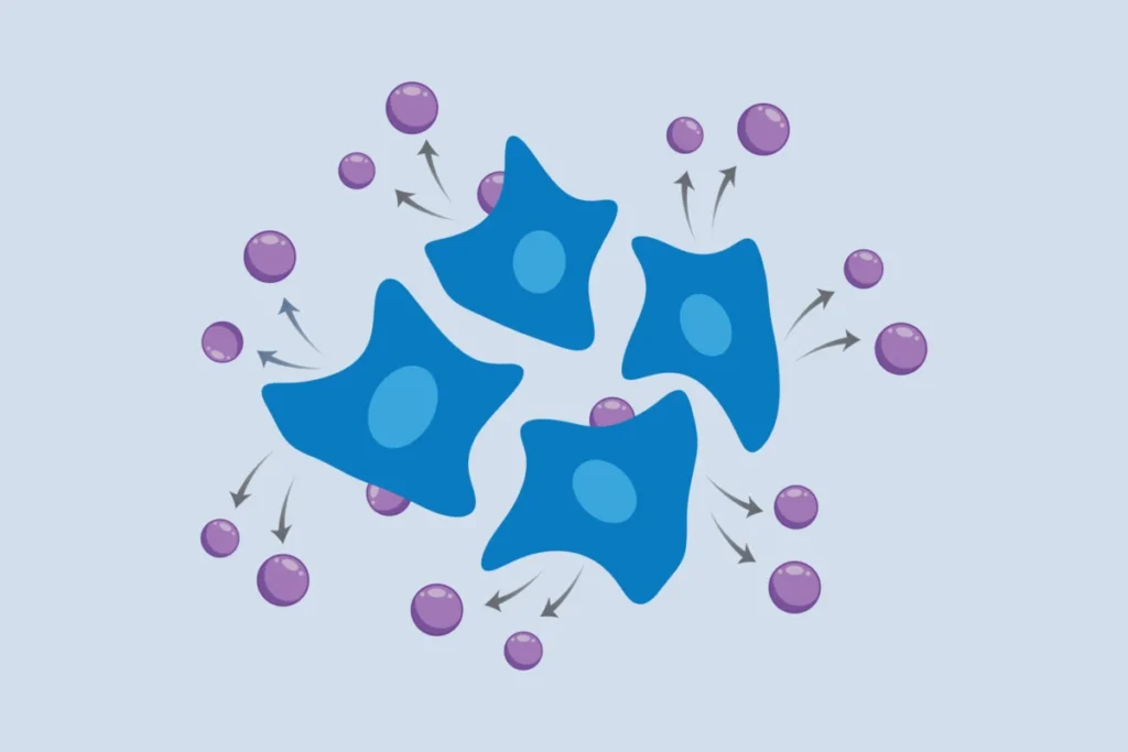
To create their non-invasive system, the researchers genetically engineered mammalian cells to express a specific retroviral structural protein. Crucially, this engineered protein possesses the remarkable ability to encapsulate not only viral RNA but also a wide array of the cell’s own messenger RNA (mRNA) and other RNA molecules. Once integrated into the cell’s membrane, this viral protein actively recruits cellular RNA, forms a protective shell around it, thereby creating a virus-like particle (VLP). These VLPs then bud off from the cell membrane and are released into the surrounding liquid culture medium, much like how viruses naturally egress from infected cells.
The beauty of this system is its simplicity and non-invasiveness. Scientists can periodically collect samples of the culture medium, isolate the RNA contained within these VLPs, and then perform standard RNA sequencing protocols. This entire process occurs without inflicting any damage or stress upon the living cells themselves. This molecularly encoded solution stands in stark contrast to more cumbersome methods involving robotics or mechanical biopsies, which often require specialized equipment and expertise, limiting their widespread adoption. As co-first author Mohamad Najia, a research fellow in the Blainey lab and the lab of George Daley at Boston Children’s Hospital, highlighted, "Compared to methods using robotics or mechanical biopsies of cells, our molecularly encoded solution could be much more broadly enabling for the average life science or biomedical lab, particularly the time dynamic questions that we hope to elucidate with this technology." This emphasis on accessibility and scalability is critical for a technology to gain widespread traction in the scientific community. Najia, along with Borrajo and co-first author Anna Le, a postdoc in the Blainey lab, played pivotal roles in bringing this vision to fruition.
Validation Across Diverse Cellular Systems and Complex Models
To rigorously test the robustness and broad applicability of their cellular self-reporting method, the research team applied it to a diverse array of cellular model systems. Their experiments successfully demonstrated the method’s efficacy in:
- Immortalized human cell lines: Common laboratory workhorses for fundamental biological research.
- Cancer cell lines: Crucial for understanding oncogenesis and testing anti-cancer therapies.
- Stem cells and neuronal cells derived from them: Enabling studies of differentiation, development, and neurological disorders.
- Primary cells from human donors: More physiologically relevant models for drug testing and disease modeling.
Furthermore, the researchers ingeniously extended the method to study co-cultures of two distinct human cell types growing together. By incorporating specific molecular tags into the virus-like particles, they were able to differentiate and analyze the transcriptional signals originating from each cell type separately. This capability is vital for understanding cell-cell interactions and the dynamics of complex cellular ecosystems.
The utility of cellular self-reporting also shines in systems with crucial three-dimensional structures that would be compromised by traditional destructive sampling. The team successfully applied their method to spheroids of human endothelial cells – three-dimensional aggregates that mimic aspects of native tissue architecture. They were able to capture short-term transcriptional changes in these spheroids following biochemical stimulation, providing dynamic insights into cellular responses within a more complex spatial context.
Perhaps one of the most compelling demonstrations of the method’s potential came through a collaboration with Professor Linda Griffith, a distinguished professor of biological and mechanical engineering at MIT, whose lab specializes in organ-on-a-chip devices. These microfluidic systems are engineered to recapitulate the physiological functions and microenvironments of human organs, offering powerful preclinical models that can minimize the need for animal testing. However, their intricate design and delicate nature make retrieving cells for analysis exceedingly difficult. The cellular self-reporting method proved to be an ideal solution. The researchers successfully monitored gene expression dynamics in endothelial cells within these organ-on-a-chip devices over time. This allowed them to uncover subtle yet significant changes in genes related to how tissues form vascular networks, revealing that these dynamics depended on the source of supporting fibroblasts (e.g., from either uterus or lung). This specific application highlights the method’s capacity to extract critical longitudinal data from complex, physiologically relevant models that were previously intractable.
Broader Impact and Transformative Implications for Biomedical Research
The development of a non-destructive, live-cell transcriptomics method carries profound implications across numerous fields of biomedical research and beyond.
Drug Discovery and Development: The pharmaceutical industry constantly seeks more predictive and efficient methods for drug screening and toxicity assessment. Current preclinical models often rely on single-time-point measurements of drug efficacy or toxicity. With cellular self-reporting, researchers can now continuously monitor how cells respond to drug candidates over hours, days, or even weeks. This enables the identification of subtle, time-dependent effects, mechanisms of drug resistance, and the kinetics of drug-induced changes in gene expression. This capability could significantly accelerate the identification of promising drug candidates, reduce attrition rates in clinical trials, and provide a more comprehensive understanding of a compound’s pharmacological profile.
Understanding Disease Pathogenesis: Many chronic diseases, such as neurodegenerative disorders, cancer, and autoimmune conditions, involve gradual changes in cellular function and gene expression over time. Traditional methods offer only fragmented views of these complex processes. This new technology allows scientists to track the precise transcriptional shifts that occur as cells transition from healthy to diseased states, or as they respond to therapeutic interventions. For example, in cancer research, it could reveal how tumor cells evolve resistance to chemotherapy in real-time. In neurobiology, it could illuminate the subtle molecular changes that precede neuronal degeneration. This longitudinal perspective is crucial for identifying early biomarkers, understanding disease mechanisms, and developing more targeted therapies.
Personalized Medicine: The ability to monitor individual patient-derived cells or organoids in response to different treatments opens new avenues for personalized medicine. Clinicians could potentially test various drug regimens on a patient’s own cells ex vivo, observing the transcriptional responses in real-time to identify the most effective and least toxic therapeutic strategy for that specific individual. This could lead to truly individualized treatment plans, moving beyond the current trial-and-error approach.
Developmental Biology and Regenerative Medicine: Understanding the precise timing and sequence of gene expression changes during cellular differentiation and tissue development is fundamental to developmental biology. This method offers an unprecedented tool to track these complex processes in live cells, providing insights into cell fate decisions, lineage commitment, and the molecular underpinnings of organogenesis. In regenerative medicine, it could be used to optimize stem cell differentiation protocols and monitor the maturation of engineered tissues.
Accessibility and Democratization of Research: The "molecularly encoded solution," as emphasized by the researchers, ensures that the method is relatively straightforward to implement in standard life science laboratories. Unlike highly specialized robotic systems or complex microdissection techniques, this approach leverages genetic engineering and standard cell culture practices, making it accessible to a broader scientific community. This democratization of advanced transcriptomic analysis is crucial for accelerating discoveries across diverse research domains.
The Broad team continues to explore new applications and biological questions that can be addressed with their system. A key future direction is to refine the approach to achieve single-cell resolution, allowing for the tracking of individual cell trajectories within a population – a capability that would represent yet another significant leap forward. For now, the researchers are optimistic that their innovative method will be widely adopted by scientists keen on understanding the dynamic intricacies of cellular life and disease.
Looking Ahead: A New Era of Dynamic Biology
The introduction of cellular self-reporting represents a pivotal moment in cellular biology. It transitions transcriptomics from a field dominated by static snapshots to one capable of dynamic, longitudinal observation. The ability to monitor gene expression in living cells, without perturbation, will undoubtedly unveil previously hidden complexities of cellular function and disease progression. As researchers across the globe begin to integrate this powerful new tool into their studies, the scientific community anticipates a new era of profound insights into fundamental biological processes and a more rapid development of innovative therapeutic strategies. The "science fiction" of a decade ago has become a reality, promising to transform our understanding of life itself.