Carnegie Mellon University’s Feinberg Lab has achieved a groundbreaking milestone in the field of tissue engineering, unveiling a novel microphysiologic system, or tissue model, constructed entirely from collagen using their innovative Freeform Reversible Embedding of Suspended Hydrogels (FRESH) 3D bioprinting technique. This advancement marks a significant departure from traditional methods that rely on synthetic materials, heralding a new era for disease research and therapeutic tissue development, particularly for conditions like Type 1 diabetes.
The Limitations of Traditional Tissue Models
For years, researchers have utilized microscopic models of human tissue to study biological processes and test potential treatments. These models, often referred to as microfluidics, organ-on-chip, or microphysiologic systems, have been indispensable tools in biomedical research. However, their construction has historically been confined to synthetic materials such as silicone rubber and plastics. This limitation arose from the technical challenges associated with precisely manipulating and structuring soft biological materials with the necessary resolution and stability.
The inherent drawback of using synthetic materials is their fundamental difference from the human body’s native environment. While these synthetic platforms can mimic certain aspects of human physiology, they often fail to fully replicate the complex biological milieu in which cells naturally reside and function. This disconnect can lead to discrepancies in cellular behavior, drug responses, and disease progression compared to what is observed in vivo. Consequently, the predictive power and translational relevance of these synthetic models have been a persistent area of concern within the scientific community.
A Paradigm Shift: Biologic, Not Synthetic
The Feinberg Lab’s breakthrough directly addresses this critical limitation by leveraging the inherent biocompatibility and biological relevance of collagen. Collagen, the most abundant protein in the human body, plays a crucial role in providing structural integrity and support to virtually all tissues and organs, including skin, bone, cartilage, and blood vessels. By constructing their microphysiologic systems entirely from collagen, cells, and other native proteins, researchers can now create models that more accurately reflect the natural biological environment.
"Now, we can build microfluidic systems in the Petri dish entirely out of collagen, cells, and other proteins, with unprecedented structural resolution and fidelity," explained Adam Feinberg, a professor of biomedical engineering and materials science & engineering at Carnegie Mellon University. "Most importantly, these models are fully biologic, which means cells function better." This statement underscores the profound implications of shifting from inert synthetic materials to dynamic, biologically active components. When cells are embedded within a matrix that closely resembles their native extracellular environment, their behavior, communication, and response to stimuli are more likely to mirror their in vivo counterparts. This enhanced biological fidelity is paramount for generating reliable research data and developing effective therapeutic strategies.
The FRESH Bioprinting Technique: Precision and Versatility
The foundation of this revolutionary approach lies in Carnegie Mellon’s novel Freeform Reversible Embedding of Suspended Hydrogels (FRESH) 3D bioprinting technique. Developed by the Feinberg lab, FRESH printing is distinguished by its ability to precisely deposit and pattern soft, living cells and complex hydrogel structures with exceptional accuracy. The technique involves embedding a sacrificial hydrogel support material within a larger bath of the same support material. Bioinks, which contain cells and other biological components like collagen, are then printed within this suspended environment. The support material can be easily dissolved or removed after printing, leaving behind the delicate, precisely structured biologic construct.
This reversible embedding process allows for the creation of intricate, three-dimensional architectures that were previously impossible to achieve with conventional bioprinting methods. Crucially, it enables the printing of hollow channels and complex vascular networks within the tissue models, a capability that is essential for mimicking the nutrient and waste exchange systems found in natural organs.
Advancing Vascularized Tissue Constructs for Diabetes Therapy
In their latest research, published in the prestigious journal Science Advances, the Feinberg lab showcases the power of their FRESH bioprinting technique by constructing more complex vascularized tissues from fully biologic materials. A key demonstration of this capability is the creation of a pancreatic-like tissue model designed to hold significant promise for treating Type 1 diabetes.
Type 1 diabetes is an autoimmune disease where the body’s immune system mistakenly attacks and destroys the insulin-producing beta cells in the pancreas. Without these cells, the body cannot produce insulin, a hormone essential for regulating blood sugar levels. Current treatments involve lifelong insulin injections and blood glucose monitoring, which can be burdensome and do not fully prevent long-term complications.
The pancreatic-like tissue developed by the Feinberg lab aims to address this by providing a bioengineered solution. The research builds upon earlier work published in Science, where the team demonstrated their foundational FRESH bioprinting capabilities. This new research significantly enhances the resolution and quality of the printed structures, enabling the creation of fluidic channels that mimic blood vessels with diameters as small as 100 microns. This level of detail is critical for ensuring that the engineered tissue can receive a sufficient supply of nutrients and oxygen, and effectively remove waste products, thereby supporting the long-term viability and function of the embedded cells.
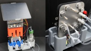
Daniel Shiwarski, assistant professor of bioengineering at the University of Pittsburgh and a former postdoctoral fellow in the Feinberg lab, elaborated on the technical advancements that made this achievement possible. "There were several key technical developments to the FRESH printing technology that enabled this work," he stated. "By implementing a single-step bioprinting fabrication process, we manufactured collagen-based perfusable CHIPS in a wide range of designs that exceed the resolution and printed fidelity of any other known bioprinting approach to date. Further, when combined with multi-material 3D bioprinting of ECM proteins, growth factors, and cell-laden bioinks and integration into a custom bioreactor platform, we were able to create a centimeter-scale pancreatic-like tissue construct capable of producing glucose-stimulated insulin release exceeding current organoid based approaches."
The mention of "glucose-stimulated insulin release" is particularly significant. This functional readout indicates that the engineered pancreatic tissue is capable of sensing glucose levels and responding by releasing insulin, a crucial hallmark of healthy pancreatic function. The comparison to "organoid based approaches" suggests that these newly developed biologic microphysiologic systems offer superior functionality and scale compared to existing organoid technologies, which are also three-dimensional cell cultures that mimic organ structure and function but may lack the vascularization and precise structural control achieved with FRESH printing.
Commercialization and Clinical Translation
The potential impact of this pioneering research extends beyond the laboratory. The technology is currently being commercialized by FluidForm Bio, a spinout company founded by Carnegie Mellon University researchers. This strategic move aims to accelerate the translation of these cutting-edge scientific discoveries into tangible clinical applications.
Dr. Andrew Hudson, Director of Tissue Therapeutics at FluidForm Bio and a co-author on the research, highlighted the company’s progress. His team has already demonstrated in an animal model that they can effectively cure Type 1 diabetes in vivo using this technology. This remarkable preclinical success provides strong evidence of the therapeutic potential of the bioengineered tissues. FluidForm Bio is now poised to advance its efforts towards human clinical trials, with plans to initiate them within the next few years. This timeline signifies a rapid progression from fundamental research to potential patient treatment, underscoring the transformative nature of the FRESH bioprinting platform.
The Power of Interdisciplinary Collaboration
Professor Feinberg emphasized the critical role of teamwork in driving such complex scientific endeavors. "It is paramount for everyone to understand the importance of team-based science in developing these technologies and the value that varied expertise, ranging from biology to materials science, brings both to the project, and our impact on society," he elaborated. This sentiment reflects the reality of modern scientific innovation, which often requires the synergistic collaboration of individuals with diverse skill sets and perspectives. The successful development of the FRESH bioprinting technique and its application in creating functional biologic tissues is a testament to the power of bringing together experts in engineering, materials science, cell biology, and medicine.
Future Directions: Beyond Construction to Optimization
Looking ahead, the focus of research is shifting from simply building complex tissues to optimizing their function and understanding how to best utilize these advanced fabrication capabilities. Feinberg articulated this forward-looking perspective: "Going forward, the question is not, can we build it? It’s more of, what do we build? The work we’re doing today is taking this advanced fabrication capability and combining it with computational modeling and machine learning, so that we can hopefully better understand what we need to print. Ultimately, we want the tissue to better mimic the disease of interest or ultimately, have the right function, so when we implant it in the body as a therapy, it’ll do exactly what we want."
This integrated approach, combining advanced 3D bioprinting with computational modeling and artificial intelligence, represents the next frontier in tissue engineering. By leveraging machine learning algorithms, researchers can analyze vast datasets to predict optimal cell compositions, growth factor concentrations, and structural designs for specific therapeutic applications. This data-driven approach promises to accelerate the design and development of more effective and personalized tissue-based therapies. The ultimate goal is to create engineered tissues that not only mimic disease states for research but also possess the precise functional characteristics required for successful implantation and long-term integration within the human body.
Open-Source Principles and Broad Adoption
In alignment with the spirit of scientific advancement and collaboration, Feinberg and his collaborators are committed to making their innovations accessible to the wider research community. They plan to release open-source designs and other technologies that facilitate broad adoption of the FRESH bioprinting platform. "We’re hoping that very quickly, other labs in the world will adopt and expand this capability to other disease and tissue areas," Feinberg added. "We see this as a base platform for building more complex and vascularized tissue systems."
This commitment to open science is crucial for accelerating progress across the entire field of regenerative medicine. By providing researchers with the tools and blueprints to replicate and build upon their work, Carnegie Mellon University is fostering a collaborative ecosystem where innovation can flourish. The FRESH bioprinting technique is envisioned not as a singular solution but as a foundational platform upon which diverse research groups can develop tailored solutions for a wide array of diseases and tissue types, from neurodegenerative disorders to cardiovascular conditions.
The implications of this research are far-reaching. The development of fully biologic, high-resolution microphysiologic systems opens up new avenues for drug discovery and development, allowing for more accurate preclinical testing of drug efficacy and toxicity. Furthermore, the ability to engineer functional tissues with unprecedented fidelity brings the prospect of regenerative medicine closer to reality, offering hope for patients suffering from a multitude of debilitating conditions. As this technology continues to mature and is adopted by researchers worldwide, it has the potential to fundamentally transform how we understand, treat, and ultimately overcome human diseases.