Epithelial cells, the ubiquitous cellular architects that form the protective linings of our bodies, have long been understood for their crucial roles in development, repair, and disease. These tiny shields constitute the outer layers of our skin, the surfaces of our organs, and the intricate networks of our blood vessels. In a developing embryo, their precise growth, division, and migration orchestrate the formation of complex biological structures. When injury strikes, be it a superficial scrape, an internal tear, or a surgical incision, epithelial cells are the first responders, migrating swiftly to the wound site to facilitate healing. Conversely, when their regulatory mechanisms fail, these same cells can undergo malignant transformation, leading to uncontrolled proliferation and the devastating spread of cancer throughout the body.
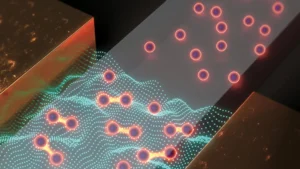
A groundbreaking study by engineers at the Massachusetts Institute of Technology (MIT) has now unveiled a previously unappreciated dimension of epithelial cell behavior: their capacity for synchronized, collective pulsing during migration. Published today in the esteemed journal Newton, the research details observations of groups of epithelial cells rhythmically moving in and out, akin to a circle of dancers drawing together and then elegantly pulling apart. This discovery adds a critical temporal layer to our understanding of cellular mechanics, moving beyond the traditional focus on spatial organization.
The MIT team meticulously measured this collective rhythmic pulsing across a spectrum of epithelial cell types, including healthy cells, cells derived from benign tumors, and those from aggressive cancerous lesions. What emerged was a startling finding: malignant epithelial cells exhibited a significantly more persistent synchronization, maintaining their collective pulse for approximately twice the duration observed in healthier cells. While the precise biological impetus behind this synchronized cellular dance remains a subject for further investigation, the researchers propose that this distinctive rhythmic behavior could serve as a vital clinical signal, offering new avenues for early detection and prognostic assessment of cancer.
Professor Ming Guo, a distinguished professor of mechanical engineering at MIT and a lead author of the study, articulated the profound implications of this observation. "More aggressive cancer cells tend to have a steadier and more persistent rhythm as compared to healthy ones," Guo stated. "We think this coordination could serve as an early warning sign of how likely a tumor is to spread. The same coordinated waves may help shape embryos during development and close wounds upon injury." This statement underscores the potential dual utility of the findings, linking fundamental developmental processes with critical disease mechanisms.
The collaborative research effort brought together a multidisciplinary team, including first author Wenhui Tang, a former MIT graduate student (SM ’20, PhD ’24), alongside Mehrana Nejad and L. Mahadevan from Harvard University, and Adrian Pegoraro of the Metrology Research Centre of the National Research Council Canada. Their combined expertise in mechanical engineering, applied mathematics, and biophysics proved instrumental in unraveling this complex cellular phenomenon.
Beyond Spatial Coordination: Unveiling Cellular Rhythms
For decades, scientific inquiry into how epithelial cells organize themselves to form complex organs and tissues has largely concentrated on spatial coordination. Researchers have primarily focused on questions pertaining to where cells move, their precise location within a growing tissue, and their ultimate destination. These spatial dynamics are undeniably crucial for understanding tissue morphogenesis and repair. However, the temporal relationship of cellular movements – how cells coordinate their actions over time – has remained a comparatively underexplored frontier.
Professor Guo’s laboratory at MIT is dedicated to investigating cell interactions with the aim of identifying patterns indicative of healthy versus diseased states. Their work often involves capturing microscopic snapshots of cells cultured in the lab, a process that allows for the identification of intriguing cellular behaviors. It was within this context that Wenhui Tang, then a member of Guo’s lab, embarked on a routine examination of a series of time-lapse movies of epithelial cells. What she began to perceive was not merely random motion, but a distinct rhythm, a recurring pattern unfolding over time.
Tang recounted her pivotal observation: "I was studying collective cell migration, and I observed cells were swelling, then squeezing together, then swelling, again and again, forming local patterns. That’s when I realized there might be something more interesting happening with these cells over time." This initial insight was the spark that ignited the extensive investigation into cellular synchronization.
The Methodology: Taking a Cellular Pulse
To systematically investigate these temporal cellular movements, the researchers meticulously designed their experiments. They began by culturing healthy, live epithelial cells in the laboratory, providing them with the necessary nutrients for natural growth, division, and movement. To enable clear visualization and differentiation of individual cells, they stained the cell nuclei with a fluorescent dye. This allowed the team to track the precise movements of each cell within the collective.
Guo elaborated on the physiological relevance of their experimental setup: "We’re looking at their natural migration process, related to how they would migrate during different processes in the body, such as when forming skin and organs, and healing wounds." This emphasis on natural migration ensures that the observed phenomena are likely reflective of in vivo processes, enhancing the study’s translational potential.
Utilizing a high-resolution confocal microscope, the team captured sequential snapshots of the cells every few minutes, extending their observation period for up up to 30 hours. The subsequent stringing together of these individual images into a time-lapse movie revealed a distinct and compelling pattern. As Tang described, "If you just stare at any one location, you can see those dots are coming together, and then going further away, then coming together again, and going further away, like waves." This visual evidence confirmed the rhythmic nature of the cellular movements.
The analysis revealed that a single pulse, encompassing the movement of cells coming together and spreading apart, typically occurred over approximately one hour. In healthy cells, this pulsing activity persisted as a slow and steady rhythm throughout the entire 30-hour observation period, indicating a robust and intrinsic cellular mechanism.

The Malignant Anomaly: Enhanced Synchronization in Cancer
Intrigued by the rhythmic behavior of healthy cells, the research team expanded their investigation to determine if other types of epithelial cells would exhibit similar synchronization. They conducted the same experiments using several different lines of human breast cancer epithelial cells, carefully selected to represent a spectrum from benign tumors to increasingly malignant forms. The observations were consistent: pockets of synchronized pulsing were evident in every cell type studied. However, a critical difference emerged when examining the most aggressive cancerous cells.
"We found the really dangerous cancer cells team up over time, and do this persistent oscillation, twice as long as healthy cells," Guo reported, highlighting the key differential. "This is unexpected. We see they really team up, synchronize, and oscillate together, which potentially facilitates their invasion." This enhanced and prolonged synchronization in malignant cells suggests a cooperative mechanism that might contribute to their aggressive behavior and metastatic potential, a process where cancer cells detach from the primary tumor and spread to distant parts of the body. Understanding the specific molecular underpinnings of this enhanced synchronization could unlock new therapeutic targets to disrupt cancer progression.
The Interplay of Density and Rhythm: Insights into Tissue Homeostasis
Beyond the differential synchronization between healthy and cancerous cells, the researchers also uncovered a fascinating correlation between cell synchronization and cell density. In each experimental dish, irrespective of the cell type, the epithelial cells continued to grow, divide, and pulse. As their numbers increased, a greater proportion of cells began to pulse together, leading to a measurable increase in overall synchronization. However, this trend was not indefinite. Once the cells reached a critical density threshold, their collective pulsing activity began to diminish.
"There’s a peak of synchrony before it decreases as cell density continues to increase," Tang explained. This observation suggests a complex regulatory mechanism where optimal synchronization occurs within a specific density range, and beyond that, cellular crowding might impede coordinated movement.
This connection between cell density and synchronization holds particular relevance for understanding various physiological and pathological conditions, notably asthma. Epithelial cells form the crucial lining of numerous organs and tissues, including the airways. In healthy individuals, these cells pack tightly together, effectively "jamming" up to form a solid, stable lining that provides a robust protective barrier for the airways. In contrast, asthmatic airways are characterized by epithelial cells that are less capable of forming this tight, jammed configuration. This structural compromise results in airways that are looser and more fragile, rendering them easily irritated and prone to inflammation, and significantly impairing their ability to heal effectively after injury or insult.
Guo and Tang posit that the observed relationship between cell density and synchronization could open new avenues for asthma research and treatment. By monitoring how potential therapeutic drugs influence the synchronization patterns of asthmatic epithelial cells, researchers might identify compounds that restore proper cell-to-cell packing and stability. This novel approach could lead to more targeted and effective treatments for asthma, moving beyond symptomatic relief to address the underlying cellular dysfunction.
Broader Implications and Future Directions: A New Frontier in Biomedicine
The discovery of synchronized cellular pulsing and its differential persistence in malignant cells carries significant implications across several fields of biomedical research.
For Cancer Diagnostics and Prognosis: The identification of a "steadier and more persistent rhythm" in aggressive cancer cells offers a novel biomarker. This cellular dance could potentially be harnessed as an early warning sign, aiding in the assessment of a tumor’s likelihood to metastasize. Future research will likely focus on developing non-invasive methods to detect and quantify this synchronization in vivo, potentially through advanced imaging techniques or biomarker assays, to translate this laboratory finding into a clinical diagnostic tool. This could refine personalized cancer treatments, allowing clinicians to tailor therapies based on a more nuanced understanding of tumor aggression.
Revolutionizing Drug Screening: The concept of disrupting cellular synchronization presents a powerful new paradigm for drug screening. As Guo envisions, "More malignant cells would be better synchronized. After treating them with a drug, if their synchronization is disrupted, then it might be an efficient drug where we can consider the next step." This offers a functional, cell-based assay that could complement existing high-throughput screening methods, accelerating the identification of compounds capable of inhibiting cancer cell invasiveness or restoring healthy epithelial function in conditions like asthma. Imagine a future where therapeutic efficacy is partly judged by a drug’s ability to ‘break the rhythm’ of cancerous cells.
Insights into Developmental Biology and Wound Healing: Professor Guo’s initial statement also highlighted the potential role of these coordinated waves in fundamental biological processes such as embryonic development and wound closure. This suggests that synchronized pulsing is not merely a pathological phenomenon but a fundamental mechanism governing tissue morphogenesis and repair. Further studies could explore how these rhythms are initiated and regulated during embryogenesis, and how their disruption might lead to developmental disorders. Similarly, understanding the dynamics of pulsing during wound healing could lead to strategies for accelerating tissue repair and minimizing scarring.
Unraveling the Molecular Mechanisms: A critical next step for researchers will be to elucidate the molecular and biophysical mechanisms that drive this synchronized pulsing. What intracellular signaling pathways are involved? How do cells communicate to achieve such coordinated movement? Are there specific genes or proteins that regulate the duration and amplitude of these pulses? Answering these questions will provide a deeper understanding of cellular mechanics and could reveal new targets for therapeutic intervention.
Expanding the Scope: Universality and In Vivo Studies: The current study primarily focused on epithelial cells cultured in vitro. Future research will need to investigate the universality of this phenomenon across different epithelial tissues and other cell types. Crucially, validating these findings in vivo – within living organisms – will be essential for their translation into clinical practice. This might involve developing advanced microscopic techniques capable of observing cellular dynamics within tissues and organs.
The implications of this discovery are far-reaching, hinting at a paradigm shift in how we understand cell behavior. By moving beyond a purely spatial view to incorporate the temporal dynamics of cellular movement, the MIT team has opened a new window into the intricate lives of our body’s protective shields. This research, supported in part by the National Institutes of Health, exemplifies the power of interdisciplinary science to uncover fundamental biological principles with profound potential for advancing human health. The rhythmic dance of epithelial cells, once an unnoticed background hum, now resonates with the promise of new diagnostic markers and therapeutic strategies for some of humanity’s most challenging diseases.