Cambridge, MA – A groundbreaking development from engineers at the Massachusetts Institute of Technology (MIT) heralds a potential revolution in cardiac care: a non-invasive pacemaker that stimulates the heart using precisely targeted ultrasound waves. This innovative design, detailed in a study published today in the prestigious journal Nature Biomedical Engineering, could one day offer a surgery-free alternative to traditional cardiac implants, dramatically improving patient outcomes and quality of life for millions worldwide.
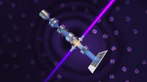
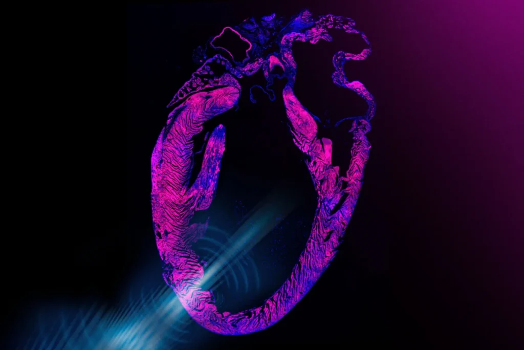
The device is conceived as a small, wearable sticker that adheres to the chest, equipped with miniature transducers that emit focused ultrasound pulses. These waves penetrate the chest cavity to interact with heart cells, triggering the opening of specific ion channels. Critically, the researchers amplified this effect through a sophisticated application of genetic engineering, making the heart cells exquisitely sensitive to the ultrasound. Once these channels open, calcium ions flood into the cells, signaling them to contract and beat, thereby regulating heart rhythm without direct electrical contact or invasive surgery.
The Enduring Challenge of Cardiac Arrhythmias
Cardiovascular diseases remain the leading cause of mortality globally, with arrhythmias – irregular heartbeats – affecting millions. In the United States alone, an estimated 3 million adults currently live with pacemakers, and countless others suffer from conditions that could benefit from cardiac rhythm management. These include bradycardia (slow heart rate), tachycardia (fast heart rate), and various forms of heart block.
For decades, the standard treatment for persistent symptomatic bradycardia and other rhythm disturbances has been the implantation of a pacemaker. These small, battery-powered devices are surgically placed under the skin in the chest, with leads extending into the heart chambers to deliver electrical impulses that maintain a regular heart rate. While undeniably life-saving and highly effective, traditional pacemakers are not without their drawbacks. The invasive nature of the procedure carries inherent risks, including infection (which can necessitate removal of the entire system), hematoma, pneumothorax, lead dislodgement or fracture, and chronic pain at the implant site. Furthermore, batteries require replacement every 7-10 years, leading to repeat surgical procedures and their associated risks. The psychological impact of living with an implanted device, though often overshadowed by its life-saving benefits, can also be significant for some patients.
"Pacemakers are one of the most important and widely used human implants, and they have saved millions of lives," acknowledges Gengxi Lu, a co-corresponding author of the paper. "But they are invasive, and they make direct contact with the beating heart. The dream for many years has been noninvasive heart stimulation with ultrasound." This MIT breakthrough aims to realize that long-held dream.
A Deeper Dive into the Mechanism: Sonogenetics at Work
The elegance of the MIT innovation lies in its dual approach: leveraging established ultrasound technology with a cutting-edge bioengineering technique known as sonogenetics. Ultrasound, a form of acoustic wave, has long been a cornerstone of medical diagnostics, safely penetrating the body to create images of organs and tissues. More recently, its therapeutic potential has been explored, with focused ultrasound being investigated for conditions ranging from Parkinson’s and Alzheimer’s disease in the brain to targeted drug delivery.
Previous studies have shown that focused ultrasound can activate heart cells, but the effect has typically been inconsistent and weak, limiting its clinical utility. The MIT team, led by Professor Xuanhe Zhao of mechanical engineering and civil and environmental engineering, sought to amplify this effect significantly. Their solution involved sonogenetics, a relatively new field inspired by optogenetics. Optogenetics involves genetically modifying specific cells to respond to light, allowing researchers to precisely control neural activity. Similarly, sonogenetics aims to engineer cells to respond robustly to sound, including ultrasound.
In their meticulous lab work, the researchers first derived human cardiac cells from embryonic stem cells. They then introduced a genetic alteration to these cells, specifically designed to increase their sensitivity to ultrasound. This manipulation resulted in the production of specialized ion channels that opened more readily when exposed to ultrasound waves.
"These channels can now ‘hear’ ultrasound better, and can open to let calcium in, which is what directly activates the cell and causes it to beat," explains Chen Gong, the paper’s first author. Calcium influx is the critical trigger for muscle contraction, including the rhythmic beating of heart cells. By genetically enhancing this natural pathway, the MIT team achieved unprecedented control over cardiac cell activity using low-intensity ultrasound.
From Lab Bench to Animal Trials: Promising Results Emerge
The efficacy of this novel approach was rigorously tested through a series of experiments. In in vitro studies, the researchers applied ultrasound waves to their engineered human cardiac cells. They observed that the pulses effectively maintained healthy and synchronized contractions, a crucial step in demonstrating the system’s ability to regulate heart rhythm.
Moving beyond the cellular level, the team then advanced to in vivo experiments using rat models. A miniature version of the pacemaker sticker was adhered to the rats’ chests. Before this, the rats received a sonogenetic, ultrasound-boosting solution, administered non-invasively, similar to a vaccine. Once the stickers were activated, the results were striking: the ultrasound quickly and effectively regulated the animals’ hearts. Rats with slow heart rates were brought up to a normal physiological rhythm, while those exhibiting irregular heartbeats were steadied, maintaining synchronization with the ultrasound’s precisely timed "ticks." These experiments provided compelling evidence that the device could quickly, safely, and non-invasively correct arrhythmias and restore normal, regular heart contractions in a living organism.
A Prototype and a Vision for Integrated Care
The current prototype developed by the team showcases the practical potential of this technology. It comprises the ultrasound sticker itself, roughly the size of a postage stamp, connected to a small, pocket-sized device containing the necessary batteries and electronics. The sticky component of the device is made from a hydrogel material, a specialty of Zhao’s group, known for its strong adhesion to skin and various materials while efficiently transmitting ultrasound waves without attenuation. The transducers embedded within the sticker can be finely tuned to generate ultrasound waves at specific frequencies, allowing for precise control over cardiac stimulation.
This research builds upon previous work by the same group, who successfully demonstrated a sticker design capable of using ultrasound to image deep organs and tissues. The ultimate vision is to combine these two capabilities into a single, integrated ultrasound sticker. This future device would not only monitor the heart’s activity in real-time but also actively regulate it, creating a truly non-invasive, closed-loop system for cardiac management.
"We believe you could one day have stickers on the body that could do long-term imaging deep in the body and also do stimulation for therapeutic effects, in a noninvasive closed-loop way," envisions Professor Xuanhe Zhao, highlighting the broader implications beyond just cardiac pacing.
The Clinical Horizon: Gene Therapy and Regulatory Pathways
Translating this pioneering technology from the lab to clinical practice involves a crucial step: the gene therapy component. For a patient to benefit from an ultrasound pacemaker, the team envisions a one-time injection, akin to a vaccine, that would deliver the genetic alteration to cardiac cells, enhancing their sensitivity to ultrasound. This would constitute a form of gene therapy, a rapidly advancing field that has seen increasing FDA approvals for conditions such as sickle cell disease, spinal muscular dystrophy, and certain inherited forms of blindness.
"We think this step would be clinically translatable as a form of gene therapy that could enable noninvasive pacemakers," Chen Gong affirms. While the prospect of gene therapy for a widespread condition like arrhythmias is exciting, it also presents a complex regulatory pathway. The FDA’s rigorous approval process for gene therapies involves extensive preclinical testing, followed by multi-phase clinical trials (Phase I for safety, Phase II for efficacy and dosing, and Phase III for large-scale efficacy and safety against standard treatments). This journey could span several years, requiring substantial investment and demonstrating long-term safety and efficacy. Key considerations will include the vector used for gene delivery (e.g., adeno-associated virus, or AAV), the specificity of its delivery to cardiac cells, the duration of gene expression, and potential immunogenicity or off-target effects.
Broader Implications: A Paradigm Shift in Bioelectronic Medicine
The development of the non-invasive ultrasound pacemaker represents a significant leap forward, not just for cardiology, but for the entire field of bioelectronic medicine. This emerging discipline seeks to modulate biological function using electrical, optical, or, in this case, acoustic signals.
The implications are far-reaching:
- Enhanced Patient Quality of Life: Eliminating surgical risks, reducing discomfort, and avoiding repeat procedures for battery changes would dramatically improve the patient experience.
- Reduced Healthcare Burden: Less invasive procedures translate to shorter hospital stays, fewer complications, and potentially lower overall healthcare costs.
- Accessibility: A non-invasive, wearable solution could make cardiac rhythm management more accessible in remote areas or developing countries where advanced surgical facilities are scarce.
- Preventative and Adaptive Care: The ability to combine monitoring and stimulation in a closed-loop system opens doors for personalized, on-demand pacing that adapts to a patient’s real-time needs, potentially even preventing severe arrhythmic events before they fully manifest.
- Beyond the Heart: As Professor Zhao notes, the underlying concept could extend to other organs. Imagine stickers for long-term imaging and therapeutic stimulation in the brain for neurological disorders, or in the gut for digestive issues, all managed non-invasively. This broader vision underscores the foundational nature of this research.
The Collaborative Effort and Future Outlook
This landmark research is the product of extensive collaboration. Professor Xuanhe Zhao and his colleagues at MIT worked closely with collaborators from Professor Qifa Zhou’s group at the University of Southern California (USC), along with researchers from Harvard University, the University of California at Los Angeles, and other groups at USC. The study’s MIT co-authors include first author Chen Gong, together with Runze Li, Won Jun Song, and former postdocs Gengxi Lu, Shucong Li, and Hsiao-Chuan Liu. Such inter-institutional and interdisciplinary cooperation is increasingly vital for tackling complex challenges in biomedical engineering.
The work received crucial financial support from various prestigious organizations, including the National Institutes of Health, the National Science Foundation, the Department of Opthamology from Research to Prevent Blindness, and the U.S. Department of War, highlighting the strategic importance of this research.
While the initial results are exceptionally promising, several steps remain on the path to clinical translation. The team is focused on further miniaturizing and integrating the device components to enhance wearability, stability, and long-term accuracy. Rigorous long-term safety and efficacy studies in larger animal models will be necessary, followed by human clinical trials. Challenges include optimizing the gene therapy delivery for sustained and targeted expression, ensuring the hydrogel’s long-term adhesion and biocompatibility, and scaling up manufacturing processes.
Nevertheless, the MIT engineers’ non-invasive ultrasound pacemaker represents a profound shift in medical technology. By merging advanced material science, precision bioengineering, and the power of sound waves, they are charting a course towards a future where life-saving interventions are less burdensome, more accessible, and truly integrated into daily life, offering a glimpse into the next generation of personalized, non-invasive medicine.