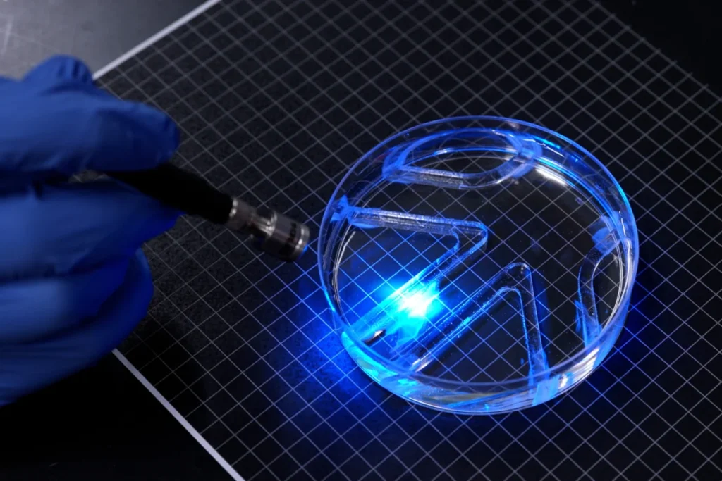
A groundbreaking development from Massachusetts Institute of Technology (MIT) engineers has redefined the capabilities of biohybrid robotics, demonstrating that even a single, paper-thin layer of muscle cells can generate sufficient power for aquatic locomotion. This innovative research, detailed in a paper published today in the journal Advanced Functional Materials, introduces a novel design for a muscle-powered swimming robot, pushing the boundaries of miniaturization and biological integration in robotic systems. The new "aquabot" represents a significant leap forward, being the first known example of a very thin, two-dimensional, muscle-powered robot capable of self-propelled movement.
The core of this aquatic marvel is a gel film, roughly the length and width of a standard stick of chewing gum, which forms the robot’s skeletal structure. This film is bifurcated into two distinct halves, each serving as a "fin." Crucially, each fin is meticulously covered with a layer of live muscle cells, engineered to be substantially thinner than a single strand of human hair. These specialized cells are genetically modified, employing a technique known as optogenetics, to twitch and contract in direct response to light stimuli. This precise control mechanism allows researchers to dictate the robot’s movements with unprecedented accuracy. By strategically shining light on one fin, its muscle layer contracts, causing the fin to flap with enough force to propel the robot through water. The ability to flash light on either fin, at varying intervals, grants full command over the swimming robot’s direction and speed.
In initial demonstrations, the paper-thin robot showcased its navigational prowess by successfully swimming and swiveling through a rudimentary watery maze. While its top speed of approximately four body lengths per minute might seem modest when compared to the sixty-five body lengths per minute achieved by Olympic swimmers, it remarkably holds its own against more leisurely aquatic inhabitants. For instance, the cow shark, known for its slow, deliberate exploration of ocean environments, navigates at a comparable pace. This comparison highlights the practical viability of the robot’s design for specific applications where speed is less critical than adaptability and the unique properties of biological actuation.
"It takes a lot of force to move through water versus air," remarked Ritu Raman, an associate professor of mechanical engineering at MIT and a lead author of the study. "The robot’s quite strong, given its size." This statement underscores the engineering triumph of generating meaningful propulsive force from such a minuscule biological engine within a viscous aquatic medium.
The Evolution of Biohybrid Robotics: Context and Precursors
The field of biohybrid robotics stands at the intersection of engineering and biology, seeking to integrate living tissues with synthetic materials to create machines with capabilities that purely artificial systems cannot replicate. These robots harness the unique attributes of biological components—such as self-healing, inherent responsiveness, and high energy efficiency—while leveraging the precision and durability of engineered structures. Historically, a significant challenge in this domain has been the construction of robust, yet agile, bio-actuated systems.
Previous efforts in biohybrid robotics, including earlier work from Raman’s group and others, often relied on bulky, three-dimensional chunks of lab-grown skeletal muscle. These constructions typically necessitate millions of cells for fabrication, leading to larger, less efficient, and more complex designs. The sheer volume of biological material required not only increases manufacturing costs and time but also limits the potential for miniaturization and delicate operations. Raman notes that thinner, less bulky designs, such as the team’s newly developed aquabot, promise to be more economical to build and inherently more efficient in their movement. This emphasis on reduced bulk and increased efficiency is a central theme in the advancement of soft robotics and bio-integrated systems.
The current breakthrough is not an isolated event but rather the culmination of a systematic research trajectory within Raman’s lab. Just last year, her group unveiled an innovative iris-inspired disk of artificial muscle tissue. In that project, a disk of gel was meticulously stamped with a pattern of concentric and radial grooves. Live muscle cells were then deposited onto this grooved surface, forming a thin layer that aligned itself along the predefined patterns. When stimulated with light, these cells exhibited coordinated movements, causing the disk to stretch and squeeze, mimicking the dilation and constriction of a human iris in response to light.
That prior work was instrumental in demonstrating the feasibility of growing muscle cells in exceptionally thin layers and arranging them in complex patterns that, when stimulated, could move in multiple, controllable directions. "People hadn’t seen this muscle architecture engineered from scratch before," Raman explained at the time. "And the cells were moving in multiple directions." However, despite the innovative architecture, the movements generated were relatively small—on the order of about 100 microns. From a practical robotics perspective, these movements were considered "tiny," insufficient to power macroscopic locomotion. This limitation served as a critical impetus for the subsequent research, which aimed to significantly amplify muscle movements to generate enough force for actual robotic propulsion.
Engineering the Optimal Skeleton: A Scientific Breakthrough in Material Design
The primary objective of the new research was to maximize the contractile force and displacement produced by the muscle cells, transforming the previously observed micron-scale twitches into movements capable of powering a swimming robot. The key to achieving this, the team discovered, lay in meticulously optimizing the "skeleton" – the underlying gel substrate upon which the muscle cells would grow and exert force.
The researchers focused on tuning three critical properties of this underlying gel: its chemical composition, its mechanical stiffness, and the precise size and shape of the grooves stamped into its surface. It is a well-established principle in tissue engineering that these parameters are fundamental "knobs" that can be adjusted to influence cell behavior and tissue development. "For engineering any type of tissue, it’s known that these are knobs you can tune," Raman stated. "And we wanted to optimize all these parameters to support live muscle cells."
In their previous iris-inspired design, the team had utilized fibrin, an ultrasoft gel. While biocompatible, fibrin proved to be problematic as it tended to quickly shrivel and deform under the contractile forces generated by the growing muscle cells. This inherent instability limited the muscle’s ability to exert maximum force and produce substantial movement. To overcome this, the researchers experimented with gelatin methacrylate (GelMA), a material widely employed in tissue engineering due to its tunable properties and biocompatibility.
Through rigorous experimentation, different formulations of GelMA were created, yielding skeletons of varying stiffnesses. The team then observed how muscle cells grew and aligned when deposited on these diverse substrates. They discovered a clear correlation: cells exhibited better alignment and, crucially, produced the most significant force when grown on stiffer GelMA gels. This finding is critical because highly aligned muscle cells tend to fuse into more robust fibers, forming stronger and more coordinated muscle tissue, which is essential for effective locomotion.
Simultaneously, the geometry of the grooves stamped into the gel surface was meticulously investigated. The team tested various groove types, from skinny square troughs to long curved valleys. Their findings indicated that muscle cells settled into optimal alignment within grooves that possessed a more square cross-section compared to curved ones. This precise architectural guidance provided by the square grooves facilitated the formation of highly organized muscle fibers, directly contributing to the enhanced force generation observed.
The thickness of the gel film also proved to be a critical parameter. After extensive testing, a half-millimeter-thin film of GelMA was identified as the optimal thickness. This specific dimension offered sufficient structural support for a single layer of muscle cells while remaining light enough to prevent the cells from peeling away from the gel during contraction. This delicate balance ensures that the muscle cells effectively transmit their contractile forces to the robot’s structure, enabling propulsion.
Finally, to further enhance the muscle’s performance, the team incorporated a "training routine." Mimicking the way physical exercise strengthens human muscles, they subjected the lab-grown muscle cells to a regimen of flashing lights. This repetitive stimulation effectively "exercised" the muscles, leading to their strengthening and improved contractile capabilities. This bio-mimetic conditioning step was instrumental in achieving the necessary force for robust robotic movement.
The culmination of these optimized parameters—the selection of GelMA, the tuning of stiffness, the precise square groove geometry, the ideal film thickness, and the muscle conditioning—resulted in a robust, aligned, and highly force-generating muscle tissue. This advanced tissue was then integrated into the two-finned robot, with the gel stamped on both sides with the optimized square-bottomed grooves and lined with the conditioned muscle cells. The cells fused into strong fibers, forming a powerful, aligned muscle tissue capable of coordinated movement.
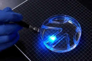
"You can think of the robot as having two independent muscles," Raman explained, highlighting the modularity of the design. "If we shine a light on just one, only that muscle moves. If shining on both, they both flap." This independent actuation is what enables precise directional control. The researchers demonstrated this by submerging the robot in a petri dish of water and manually maneuvering a light source over it. The robot faithfully followed the light, flapping its fins in response to navigate through a small maze placed within the dish, unequivocally demonstrating its capability for directed locomotion.
Broader Implications and Future Horizons
This pioneering work by MIT engineers holds profound implications for the future of robotics and biomedical engineering. The development of a thin, efficient, and biologically powered swimming robot opens doors to a new generation of adaptable and resilient machines.
One of the most immediate and exciting applications lies in exploration. Biohybrid robots, with their soft, compliant, and responsive living tissues, are uniquely suited for exploring environments that are too fragile, unpredictable, or hazardous for conventional, rigid hardware. Imagine these bots delicately navigating pristine coral reefs without causing damage, or exploring microscopic biological systems within an organism without disrupting its delicate balance. They could also be deployed in environments contaminated by hazardous chemicals, where their biological components might offer a degree of resilience or even self-repair capabilities that synthetic materials lack. The ability of living tissue to respond to its surroundings and, critically, to heal itself, provides a distinct advantage over purely mechanical systems, which often fail irrevocably upon damage.
Beyond environmental exploration, the technology has significant potential in biomedical devices. Micro-surgical robots capable of navigating the intricate pathways of the human body, delivering drugs with pinpoint accuracy, or performing diagnostics in real-time could revolutionize medicine. Their biocompatibility and inherent softness would minimize tissue damage, offering a less invasive alternative to current procedures. Similarly, these robots could serve as sophisticated environmental monitoring sensors in aquatic ecosystems, offering insights into water quality, detecting pollutants, or tracking the movement of microorganisms, all with minimal ecological footprint.
From an economic and fabrication standpoint, the shift towards thinner, less bulky designs is a critical advancement. Traditional biohybrid robots, requiring vast quantities of cells and complex three-dimensional scaffolding, are expensive and time-consuming to produce. The streamlined design of the MIT aquabot, utilizing a single, thin layer of muscle cells, suggests a pathway toward cheaper, more scalable manufacturing processes, making biohybrid robotics more accessible for diverse applications.
However, challenges remain. The lifespan and maintenance of living tissue within an autonomous robot present significant hurdles. Living cells require a constant supply of nutrients, efficient waste removal, and a precisely controlled environment to thrive. Integrating these life-support systems into a miniature, self-contained robot operating autonomously in varied environments will require further innovation. Scalability is another area for future research; can this technology be effectively scaled up for larger tasks or scaled down even further for nanorobotics? Furthermore, the integration with more sophisticated control systems beyond simple light patterns, perhaps incorporating electrical or chemical stimuli, could unlock more complex behaviors and autonomous decision-making capabilities. The ethical considerations surrounding biohybrid organisms, while perhaps less contentious than full animal-human hybrids, will also require ongoing dialogue as the field progresses.
The interdisciplinary nature of this research, blending mechanical engineering, materials science, biology, and genetic engineering, underscores the collaborative spirit driving modern scientific discovery. By drawing inspiration from biology and leveraging advanced engineering techniques, Raman’s team is not just building robots; they are laying the groundwork for a new paradigm of "living" machines – systems that can adapt, respond, and even heal, much like the organisms that inspired their creation. This work brings closer the vision of soft, responsive, and self-healing biohybrid robots that could one day perform delicate jobs in environments too fragile or unpredictable for conventional hardware. The implications extend beyond mere utility, inviting us to reconsider the very definition of a machine and its potential role in shaping our future.