CAMBRIDGE, MA – Scientists at the Broad Institute of MIT and Harvard, in a groundbreaking advance published in the prestigious journal Cell, have unveiled a novel "cellular self-reporting" method that allows researchers to monitor the genetic activity of living cells without the need for destructive sampling. This innovative technique overcomes a long-standing limitation in cell biology by enabling the repeated analysis of a cell’s transcriptome—the complete set of RNA molecules—from the surrounding culture medium, providing unprecedented insights into dynamic cellular processes over time.
For decades, understanding the intricate workings of a cell has hinged significantly on analyzing its transcriptome. The transcriptome serves as a dynamic blueprint, reflecting which genes are actively being expressed and at what levels, thereby dictating a cell’s identity, function, and response to its environment. Traditional methods for capturing this crucial information, primarily RNA sequencing, have invariably required the physical destruction of the cells. While highly powerful for generating snapshots of gene expression at a single point in time, these destructive techniques inherently preclude the study of how gene activity evolves within the same population of cells as they mature, differentiate, respond to drugs, or succumb to disease progression. This limitation has created a significant hurdle in fields ranging from developmental biology to drug discovery and personalized medicine, where the temporal dimension of cellular behavior is paramount.
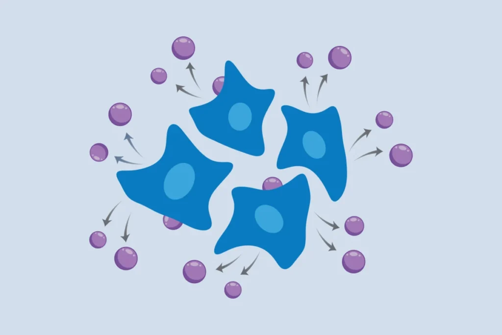
The newly developed approach, spearheaded by researchers at the Broad Institute and MIT, fundamentally shifts this paradigm. Instead of lysing cells to extract their RNA, the method leverages engineered cells to spontaneously package and release their RNA into the surrounding culture medium via specialized virus-like particles (VLPs). Scientists can then simply collect samples of this medium, isolate the secreted RNA, and perform sequencing to obtain a comprehensive profile of the cells’ transcriptional state. The non-invasive nature of this "self-reporting" mechanism means that the same cell population can be monitored repeatedly, offering a dynamic, longitudinal view of gene activity that was previously unattainable. This capability promises to unlock deeper understanding of cellular trajectories, disease mechanisms, and drug efficacy, moving beyond static observations to capture the true fluidity of biological systems.
A Decade-Long Quest to Revolutionize Transcriptomics
The journey to this transformative discovery began more than a decade ago in the laboratory of Paul Blainey, a core member of the Broad Institute and a professor of biological engineering at MIT, and the senior author of the study. Blainey vividly recalls the rudimentary nature of existing RNA sequencing methods at the time, describing them as "a bit medieval" due to their reliance on physically disrupting cells. This dissatisfaction fueled a long-term commitment to developing a non-destructive alternative.
The inspiration for a molecularly encoded solution drew from the remarkable efficiency and broad adoption of technologies like CRISPR-based gene editing, which demonstrated the power of elegant molecular tools to democratize complex biological investigations. Blainey, alongside study first author Jacob Borrajo, recognized that while a molecular approach would be inherently challenging and time-consuming to develop, its eventual scalability and ease of use by other laboratories would far outweigh the initial investment. This foresight laid the groundwork for a sustained research effort, characterized by high-risk, high-reward thinking, which Blainey now sees as a profound validation of that initial vision. "Our lab focuses our time and resources on developing tools that will actually get used and make real impact on the broader field," Blainey stated, adding, "It’s so gratifying to see a real coming to fruition of this concept, which was complete science fiction when we started. It’s a great example of the innovative impact long-term high-risk, high-reward research can have."
The critical breakthrough came from an unexpected source: retroviruses. These viruses, through millions of years of evolution, have perfected the ability to encapsulate their RNA genomes within protein shells, facilitating their spread from one host cell to another. The research team ingeniously co-opted this natural mechanism. They engineered mammalian cells to express a specific retroviral structural protein. Crucially, this protein was modified to not only package viral RNA but also to efficiently encapsulate a cell’s own messenger RNA (mRNA) and other RNA species. Once integrated into the cell’s membrane, this viral protein recruits cellular RNA, forms a protective virus-like particle around it, and then buds off from the cell surface, releasing these RNA-laden particles into the surrounding culture medium. This elegant "special delivery" system allows for the continuous secretion of a cell’s transcriptional information without causing any harm to the cell itself.
Broad Applicability and Experimental Validation
The immediate utility and versatility of this cellular self-reporting method were rigorously demonstrated across a diverse range of cellular model systems, underscoring its potential to become a cornerstone technology in biological research. The researchers successfully applied the technique to immortalized human cell lines, which are foundational tools in countless laboratories worldwide. Furthermore, they validated its performance in various cancer cell lines, opening avenues for real-time monitoring of tumor progression, drug resistance development, and therapeutic responses.
Beyond established cell lines, the method proved effective in more complex and physiologically relevant systems, including human induced pluripotent stem cells (iPSCs) and the neuronal cells differentiated from them. This capability is particularly significant for studying developmental processes, neurological disorders, and regenerative medicine, where the dynamic changes in gene expression during differentiation and maturation are critical to understand. The method also performed robustly with primary cells directly isolated from human donors, affirming its potential for clinical and translational research.
To showcase its capacity for complex multicellular environments, the team conducted experiments with co-cultures of two distinct human cell types growing together. By incorporating unique tags into the virus-like particles produced by each cell type, they were able to differentiate and independently analyze the transcriptional signals emanating from each population. This feature is invaluable for studying cell-cell interactions, tissue microenvironments, and the intricate communication networks that govern biological systems.
A particularly compelling application involved studying systems with crucial three-dimensional structures, which are often challenging to analyze with traditional destructive methods. The researchers successfully applied their self-reporting technique to spheroids of human endothelial cells, capturing short-term transcriptional changes following biochemical stimulation. Spheroids, being 3D aggregates of cells, more closely mimic in vivo tissue architecture than conventional 2D cell cultures, and the ability to monitor their gene expression non-invasively represents a significant leap forward.
Perhaps one of the most exciting demonstrations involved a collaboration with Linda Griffith, a professor of biological and mechanical engineering at MIT, known for her pioneering work on organ-on-a-chip devices. These microfluidic platforms represent miniature, functional models of human organs, designed to mimic the complex physiology of tissues and organs in vitro. Organ-on-a-chip technology holds immense promise for reducing reliance on animal models and accelerating drug development, but the inherent complexity of these devices often makes retrieving cells for analysis extremely difficult, if not impossible, without compromising the model’s integrity.
With cellular self-reporting, the Broad and MIT team monitored gene expression dynamics in endothelial cells residing within these sophisticated organ-on-a-chip devices over extended periods. This enabled them to reveal dynamic changes in genes associated with the formation of vascular networks (angiogenesis). Crucially, their findings demonstrated that these vascularization patterns were dependent upon the source of supporting fibroblasts—whether they originated from the uterus or the lung. This level of dynamic, context-dependent transcriptional insight from an intact 3D organoid system marks a significant milestone, illustrating the method’s power to unravel subtle yet critical biological nuances in highly relevant physiological models.
Mohamad Najia, a co-first author of the study and a research fellow in the Blainey lab and the lab of George Daley at Boston Children’s Hospital, highlighted the practical advantages: "Compared to methods using robotics or mechanical biopsies of cells, our molecularly encoded solution could be much more broadly enabling for the average life science or biomedical lab, particularly the time dynamic questions that we hope to elucidate with this technology." Najia led the work alongside Jacob Borrajo and co-first author Anna Le, a postdoc in the Blainey lab, underscoring the collaborative effort behind the innovation.
Transformative Implications for Biomedical Research
The advent of cellular self-reporting stands to profoundly impact numerous facets of biomedical research and drug development, offering a dynamic lens into cellular behavior that was previously obscured.
Drug Discovery and Development: The ability to monitor gene expression in real-time within living cells and organoids can revolutionize drug screening. Researchers can now observe how potential drug candidates alter cellular transcriptional programs over time, identify early markers of efficacy or toxicity, and understand mechanisms of action or resistance without destroying valuable samples. This could significantly accelerate the identification of promising compounds, reduce attrition rates in preclinical development, and facilitate the development of more targeted therapies. For instance, in oncology, researchers could track how cancer cells adapt to chemotherapy, identifying genes upregulated in resistance pathways as they emerge.
Disease Modeling and Progression: Understanding the temporal dynamics of disease is critical. Whether it’s the gradual accumulation of cellular dysfunction in neurodegenerative diseases like Alzheimer’s and Parkinson’s, the dynamic changes during inflammatory responses, or the subtle shifts leading to metabolic disorders, cellular self-reporting offers an unparalleled tool. Researchers can now track the transcriptional evolution of diseased cells in vitro, gaining insights into disease onset, progression, and potential intervention points. This could lead to the identification of new biomarkers for early diagnosis and therapeutic targets.
Personalized Medicine: The method holds immense promise for personalized medicine. By culturing patient-derived cells or organoids and exposing them to various treatments, clinicians could potentially monitor an individual’s unique transcriptional response in real-time. This could inform treatment selection, optimize dosing, and predict patient outcomes with greater precision, moving towards truly individualized therapeutic strategies.
Fundamental Cell Biology: Beyond translational applications, cellular self-reporting will empower basic scientists to address fundamental questions about cellular identity, differentiation, and environmental responses. How do stem cells precisely orchestrate gene expression changes during differentiation into specialized tissues? What are the immediate and long-term transcriptional consequences of specific environmental stimuli, nutrient deprivation, or pathogen exposure? These are questions that can now be explored with unprecedented temporal resolution.
Overcoming Current Research Bottlenecks: The non-destructive nature of the method also addresses practical challenges. For rare cell types or precious patient samples, where destructive sampling is prohibitive, cellular self-reporting offers a viable path to gain rich molecular insights. Furthermore, its molecularly encoded nature makes it amenable to high-throughput applications, allowing for the simultaneous monitoring of numerous conditions or cell lines, a significant advantage over labor-intensive robotic or mechanical methods.
Future Directions and Broader Adoption
The Broad team is actively pursuing new applications and biological questions that can be addressed with their innovative system. A key area of ongoing research involves refining the approach to make it feasible for studying single cells. While the current method provides population-level transcriptomic data, achieving single-cell resolution in a non-destructive, longitudinal manner would represent another quantum leap in biological understanding. The complexities of capturing the minute RNA output from a single cell without damaging it are substantial, but the groundwork laid by this current breakthrough provides a strong foundation.
For now, the researchers are optimistic that scientists across various disciplines will embrace cellular self-reporting to tackle dynamic biological questions. The simplicity of sampling the culture medium, coupled with the power of longitudinal data, positions this method as a versatile and broadly applicable tool for any lab interested in understanding how cells and tissues change over time. From observing the subtle shifts in gene expression during embryonic development to tracking the molecular battle against infectious agents, the "message in a bottle" delivered by living cells promises to unlock a new era of dynamic biological discovery.