Cambridge, MA – Scientists at the Broad Institute of MIT and Harvard, in collaboration with researchers at MIT, have introduced a groundbreaking "cellular self-reporting" method that enables the continuous monitoring of a living cell’s transcriptome without causing its destruction. This innovative approach, detailed in a recent publication in the prestigious journal Cell, represents a significant paradigm shift in molecular biology, moving beyond the traditional one-time snapshot of cellular genetic activity to offer dynamic, real-time insights into how cells function, mature, and respond to various stimuli over extended periods.
For decades, the study of the transcriptome – the complete set of RNA molecules produced by a cell – has been fundamental to understanding cellular identity, function, and disease mechanisms. Conventional methods, such as bulk RNA sequencing and more recently single-cell RNA sequencing, provide invaluable data by measuring all the RNA present within a cell. However, a critical limitation has persisted: these techniques necessitate the lysis, or destruction, of the cell to access its RNA content. This inherent destructive nature means that researchers could only obtain a static snapshot of a cell’s transcriptional state at a single point in time, precluding the observation of dynamic changes within the same cell population or individual cells as they evolve, differentiate, or react to environmental shifts. The new method bypasses this fundamental hurdle, allowing scientists to repeatedly sample and analyze the RNA released by living cells, thus opening up entirely new avenues for biological discovery and therapeutic development.
The Paradigm Shift: Moving Beyond the Snapshot
The inability to track gene expression dynamics in living cells has long been a bottleneck in numerous fields of biological research. In developmental biology, understanding how cells differentiate from stem cells into specialized tissues involves a complex cascade of gene activation and repression. In immunology, the response of immune cells to pathogens or therapeutic interventions is a highly dynamic process. In oncology, tracking the transcriptional evolution of cancer cells as they develop resistance to drugs or metastasize is crucial for effective treatment strategies. Neurodegenerative diseases, characterized by subtle, progressive changes in neuronal gene expression, have also been particularly challenging to study longitudinally at the molecular level.
Existing technologies, while powerful, inherently sacrifice the temporal dimension. Researchers would typically have to take multiple samples at different time points, each requiring a fresh batch of cells and yielding a new set of "snapshots." Comparing these distinct snapshots across different cell populations introduced variability and made it difficult to discern true dynamic changes from inter-sample differences. The "cellular self-reporting" method directly addresses this critical unmet need, offering a continuous narrative of gene activity rather than a series of disconnected images.
A Decade in the Making: The Genesis of Cellular Self-Reporting
The journey to this breakthrough began more than a decade ago within the laboratory of Paul Blainey, a core member of the Broad Institute and a professor of biological engineering at MIT. Blainey and his team embarked on a high-risk, high-reward endeavor to develop a non-destructive method for RNA sequencing, driven by the recognition that existing techniques were, in his words, "a bit medieval and involved stabbing cells or cutting pieces off of them."
The initial challenge was formidable. How could one access the delicate RNA molecules within a cell without compromising its viability or integrity? The inspiration for a molecular solution emerged from an unexpected source: retroviruses. These ancient biological entities have evolved over millions of years to efficiently package their RNA genomes into protective protein shells, known as virus-like particles (VLPs), which they then release to spread infection from one cell to another. This natural mechanism of molecular self-packaging and delivery provided a blueprint for the Broad and MIT team.
Led by Blainey and with significant contributions from study co-first authors Jacob Borrajo, Mohamad Najia (a research fellow in the Blainey lab and the lab of George Daley at Boston Children’s Hospital), and Anna Le (a postdoc in the Blainey lab), the team committed to developing a purely molecular method. This commitment was crucial, as a molecularly encoded solution promised scalability and ease of adoption by other laboratories, akin to the widespread impact of CRISPR-based technologies. "Our lab focuses our time and resources on developing tools that will actually get used and make real impact on the broader field," stated Paul Blainey. "It’s so gratifying to see a real coming to fruition of this concept, which was complete science fiction when we started. It’s a great example of the innovative impact long-term high-risk, high-reward research can have."
Mechanism Unveiled: How Cells "Self-Report"
The core of the "cellular self-reporting" technology lies in ingeniously repurposing a retroviral mechanism. The researchers engineered mammalian cells to express a specific retroviral structural protein. This protein, once integrated into the cell’s membrane, possesses the remarkable ability to recruit and encapsulate not only viral RNA but also the cell’s own diverse RNA molecules.
Here’s a step-by-step breakdown of the process:
- Engineering Cells: Mammalian cells are genetically engineered to express a specific retroviral structural protein. This protein is typically involved in forming the viral capsid.
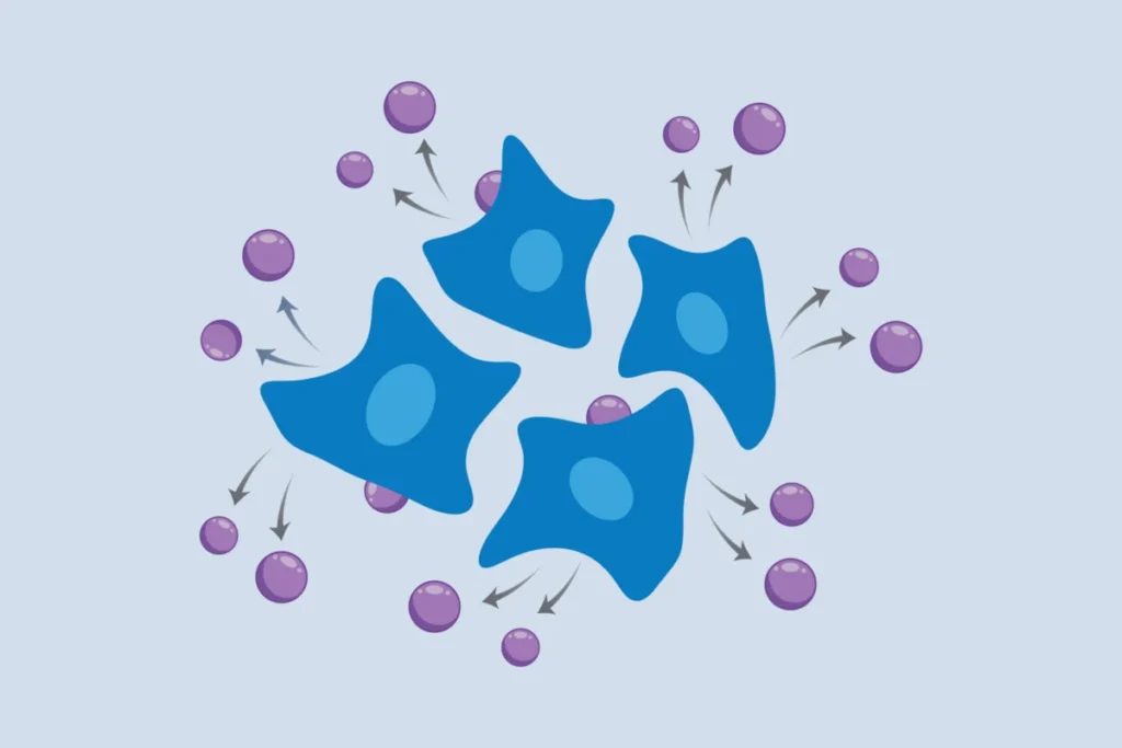
- RNA Encapsulation: Once expressed, this viral protein localizes to the cell membrane. It then begins to recognize and encapsulate various cellular RNA molecules, forming a protective protein shell around them. This creates a non-infectious, self-assembled virus-like particle (VLP) loaded with cellular RNA.
- Budding and Release: These RNA-laden VLPs then bud off from the cell membrane and are released into the surrounding culture medium, much like how actual viruses would exit an infected cell.
- Sampling and Analysis: Scientists can then simply collect a sample of the culture medium. From this medium, the released VLPs are isolated, their RNA cargo extracted, and subsequently sequenced using standard RNA sequencing protocols.
Crucially, this entire process occurs without disturbing the living cells themselves. The cells continue to grow, divide, and function normally, providing a continuous stream of their transcriptional information. This elegant molecular solution stands in stark contrast to previous methods that relied on robotics, microfluidic devices, or mechanical biopsies, which often introduced stress or damage to the cells and were far more complex to scale. As Mohamad Najia explained, "Compared to methods using robotics or mechanical biopsies of cells, our molecularly encoded solution could be much more broadly enabling for the average life science or biomedical lab, particularly the time dynamic questions that we hope to elucidate with this technology."
Validation Across Diverse Biological Systems
To demonstrate the broad applicability and robustness of their new method, the Broad and MIT team rigorously tested it across a wide array of cellular models. Their findings confirmed its efficacy in:
- Immortalized Human Cells: Common laboratory cell lines, demonstrating basic functionality.
- Cancer Cell Lines: Providing a means to study the dynamic gene expression changes associated with cancer progression and response to therapies.
- Stem Cells and Neuronal Cells: Highlighting its potential in developmental biology and neurobiology, where cell differentiation and maturation are critical processes involving significant transcriptional shifts.
- Primary Cells from Human Donors: Underscoring its relevance for translational research and patient-specific studies.
The researchers also pushed the boundaries by applying the method to more complex systems. In one experiment, they co-cultured two different human cell types. By incorporating distinct molecular tags onto the virus-like particles released by each cell type, they were able to differentiate and analyze the transcriptome signals from both populations simultaneously, providing insights into intercellular communication and interaction dynamics.
Furthermore, the method proved invaluable for studying systems with crucial three-dimensional structures that are highly sensitive to disturbance. The team successfully applied cellular self-reporting to spheroids of human endothelial cells – 3D cell aggregates that mimic aspects of tissue architecture. They were able to capture rapid transcriptional changes in these spheroids following biochemical stimulation, demonstrating the method’s ability to monitor subtle, short-term responses in a physiologically relevant context.
A particularly impactful application involved a collaboration with Linda Griffith, a professor of biological and mechanical engineering at MIT, known for her pioneering work on organ-on-a-chip devices. These microfluidic systems are engineered to mimic the complex physiology and function of human organs, offering powerful platforms for disease modeling and drug testing, often minimizing the need for preclinical animal studies. However, the intricate architecture of organ-on-a-chip devices makes it exceedingly difficult to retrieve cells for traditional destructive analyses. With cellular self-reporting, the researchers could continuously monitor gene expression dynamics in endothelial cells within these devices over time. This allowed them to uncover nuanced changes in genes related to the formation of vascular networks, revealing how these processes were influenced by the source of supporting fibroblasts (e.g., from uterus or lung tissue). This integration marks a significant step towards leveraging organ-on-a-chip technology for dynamic, high-throughput molecular profiling.
Broader Impact and Implications for Research and Medicine
The introduction of cellular self-reporting stands to revolutionize several key areas of biomedical research and has profound implications for drug discovery and personalized medicine.
1. Drug Discovery and Development:
The ability to monitor gene expression in living cells over time provides an unprecedented tool for drug screening and toxicity assessment. Researchers can now:
- Track Drug Efficacy: Observe how potential drug candidates modulate gene expression pathways in real-time, providing a clearer picture of their mechanism of action and dose-dependent effects.
- Assess Toxicity: Identify early transcriptional signatures of cellular stress or damage induced by compounds, potentially flagging toxic candidates much earlier in the development pipeline.
- Identify Biomarkers: Discover dynamic gene expression patterns that serve as biomarkers for drug response, resistance, or disease progression, aiding in patient stratification for clinical trials.
- Reduce Animal Testing: By integrating with organ-on-a-chip models, this method can significantly enhance the predictive power of in vitro systems, potentially reducing the reliance on animal models and accelerating the preclinical development phase. The global pharmaceutical market, projected to reach over $2 trillion by 2028, constantly seeks methods to reduce the exorbitant costs and lengthy timelines associated with drug development, making this innovation particularly valuable.
2. Understanding Disease Mechanisms:
Many diseases, from cancer to neurodegeneration, are characterized by dynamic changes in cellular state. This method allows scientists to:
- Map Disease Progression: Chart the molecular trajectory of disease development, identifying key transcriptional shifts that drive pathogenesis. This is especially critical for chronic conditions where subtle changes accumulate over time.
- Investigate Cellular Differentiation and Reprogramming: Gain deeper insights into the intricate gene regulatory networks that govern cell fate decisions, crucial for regenerative medicine and understanding developmental disorders.
- Study Immune Responses: Monitor the evolving gene expression profiles of immune cells during infection, inflammation, or autoimmune reactions, providing a clearer understanding of immune system dynamics.
3. Personalized Medicine:
In the era of personalized medicine, understanding individual patient responses is paramount. The ability to dynamically profile patient-derived cells (e.g., from biopsies or induced pluripotent stem cells) in vitro could lead to:
- Optimized Therapeutic Strategies: Tailoring treatments based on how a patient’s cells respond to various drugs over time, potentially leading to more effective and less toxic therapies.
- Prognostic Tools: Developing new diagnostic and prognostic tools by identifying dynamic molecular signatures that predict disease outcome or treatment success.
The global transcriptomics market, valued at approximately $7-8 billion in 2023, is projected to grow substantially, reaching upwards of $20 billion by 2030. This growth is largely fueled by continuous technological advancements in sequencing and a burgeoning demand for detailed gene expression analysis across research and clinical applications. The Broad Institute’s innovation directly addresses a significant unmet need within this rapidly expanding landscape, promising to drive further expansion and unlock previously inaccessible biological insights.
Expert Perspectives and Future Directions
The scientific community is poised to embrace this new technology with considerable enthusiasm. The scalability and molecular elegance of the cellular self-reporting method make it highly accessible, a key factor for widespread adoption. As Paul Blainey emphasized, the goal was always to create tools that would have a "real impact on the broader field." This innovation undeniably fulfills that vision.
While the current method focuses on population-level analysis, the Broad team is already looking ahead. One of their primary goals is to refine the approach to achieve single-cell resolution. The ability to track the transcriptome of individual living cells over time would represent another monumental leap, offering unparalleled insights into cellular heterogeneity and individual cell fate decisions. Further down the line, researchers might explore the feasibility of adapting similar principles for in vivo applications, though the challenges associated with delivering and retrieving VLPs within a living organism would be significantly more complex.
For now, the immediate call to action from the Broad team is for scientists interested in understanding how cells and tissues change over time to integrate this novel method into their research. It offers a powerful new lens through which to observe the dynamic choreography of gene expression, promising to unravel long-standing biological mysteries and accelerate the development of next-generation therapies. This "message in a bottle" from living cells heralds a new era in molecular biology, where the once-static picture of the transcriptome gives way to a vibrant, unfolding narrative of life itself.