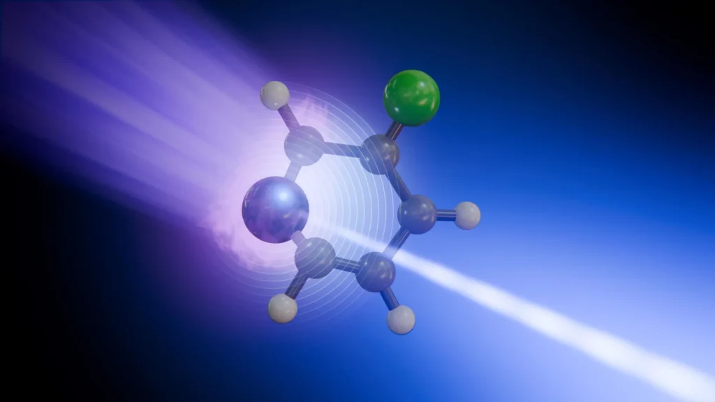
In a landmark achievement for the field of ultrafast photochemistry, an international team of researchers has successfully mapped the internal energy pathways of a molecule with unprecedented atomic-level resolution. By utilizing the ultra-rapid X-ray flashes generated at the European XFEL (X-ray Free-Electron Laser) facility, the scientists were able to observe how energy redistributes itself within a single molecule in the trillionths of a second following light absorption. This breakthrough reveals a surprising complexity: individual atoms within the same molecule serve as distinct "reporters," each capturing different facets of the molecule’s rapid structural and electronic transformation.
The study, which focused on the heterocyclic molecule 3-fluoropyridine, marks a significant leap forward in our ability to witness the fundamental "flickers" of chemical reactions. For decades, scientists have sought to understand the exact mechanisms by which matter processes energy from light—a process that governs everything from human vision and photosynthesis to the degradation of DNA under ultraviolet radiation. Until now, capturing these movements with both high temporal and spatial precision simultaneously has remained a significant challenge.
The Molecular Subject: 3-Fluoropyridine
The researchers chose 3-fluoropyridine as their subject due to its specific structural properties. A small, ring-shaped molecule, it consists of five carbon atoms, one nitrogen atom, and one fluorine atom. This arrangement makes it an ideal candidate for multi-site sensitivity studies. Because nitrogen and fluorine are located at different positions within the ring and possess different electronic environments, they provide unique vantage points for observing how the molecule reacts when "kicked" by a pulse of energy.
In its natural state, 3-fluoropyridine maintains a flat, planar structure. However, this stability is disrupted the moment it absorbs ultraviolet (UV) light. The absorption of a UV photon pushes the molecule’s electrons into an excited state, initiating a cascade of movements as the molecule attempts to shed this excess energy and return to its stable ground state.
The Mechanics of a Molecular Transformation
The transformation begins with a femtosecond-scale (one quadrillionth of a second) event. When the UV laser pulse hits the 3-fluoropyridine molecule, the electronic distribution shifts instantaneously. This change in electronic "cloud" density alters the forces acting on the atoms’ nuclei, causing the molecule to lose its flat shape and begin to bend and twist.
As the molecule distorts, it reaches a critical juncture known as a "conical intersection." In the world of quantum chemistry, conical intersections are the "funnels" of molecular energy. They are short-lived regions where the energy levels of the excited state and the ground state meet. At these points, the motion of the electrons and the motion of the atomic nuclei become "strongly coupled," meaning they influence each other so intensely that they can no longer be treated as separate entities.
The conical intersection acts as a high-speed bypass, allowing the molecule to transition from an electronically excited state back to its ground state almost instantly. However, the energy doesn’t simply vanish; the electronic energy is converted into "vibrational energy." The molecule, now back in its ground state, begins to vibrate violently, like a plucked string, as the energy ripples through its bonds.
Utilizing the European XFEL for Real-Time Observation
To capture this process, which unfolds over the course of just a few picoseconds (trillionths of a second), the research team employed a sophisticated technique known as time-resolved X-ray photoelectron spectroscopy (tr-XPS). This experiment was conducted at the Small Quantum Systems (SQS) instrument at the European XFEL, a 3.4-kilometer-long facility that generates X-ray pulses so short and bright they can act as a high-speed camera for the subatomic world.
The experimental setup followed a "pump-probe" methodology:
- The Pump: A brief ultraviolet laser pulse (the pump) was fired at a stream of 3-fluoropyridine molecules to initiate the energy absorption and structural bending.
- The Probe: A precisely timed soft X-ray pulse (the probe) followed the UV pulse. This X-ray pulse had enough energy to knock "deeply bound" electrons out of their orbits near the nuclei of either the nitrogen or the fluorine atoms.
- The Detection: By measuring the kinetic energy of these released electrons, the researchers could determine the "binding energy" of the atoms at that exact moment. Because an atom’s binding energy is sensitive to its local chemical environment, any change in the molecule’s shape or electron distribution was reflected in the data.
By repeating this process thousands of times and varying the delay between the UV "pump" and the X-ray "probe" by increments of just a few femtoseconds, the team was able to stitch together a frame-by-frame movie of the molecule’s transformation.
Divergent Stories: Nitrogen vs. Fluorine
The most striking finding of the study was that the nitrogen and fluorine atoms provided entirely different accounts of the energy transfer. The fluorine atom, located further from the primary site of electronic excitation, acted as a clean indicator of vibrational relaxation. Its signal primarily tracked how the whole molecule’s "shaking" subsided as the energy dissipated into the environment.
The nitrogen atom, however, told a much more intricate story. Because the nitrogen atom is more intimately involved in the initial electronic excitation and the subsequent passage through the conical intersection, its signal was a hybrid. It reflected the rapid redistribution of electrons in the first few hundred femtoseconds, followed by the structural distortion of the ring, and finally the vibrational cooling.
"We can now see that not every atomic site tells the same story in the signals we capture from our X-ray pulses," explained Antonio Picón, a co-author of the study from the Instituto de Ciencia de Materiales de Madrid (ICMM-CSIC). "Some atoms report where the charge is going, while others reveal how the whole molecule vibrates."
This "multi-site sensitivity" is revolutionary because it allows chemists to stop looking at molecules as monolithic blocks and start seeing them as dynamic systems where different parts perform different roles during a reaction.
Chronology of the Transformation
Based on the data collected at the European XFEL and supported by advanced computer simulations, the timeline of the 3-fluoropyridine transformation can be broken down into four distinct phases:
- 0 to 50 Femtoseconds: The UV pulse hits the molecule. Electrons are promoted to a higher energy state, and the electronic cloud begins to shift away from the nitrogen atom.
- 50 to 300 Femtoseconds: The molecule begins to lose its planar shape. It reaches the conical intersection. Here, the electronic energy starts "leaking" into the movement of the atoms.
- 300 to 1,000 Femtoseconds (1 Picosecond): The molecule crosses the conical intersection and returns to the ground state. The electronic energy is now fully converted into internal heat (vibrations).
- 1 to 5 Picoseconds: The molecule undergoes vibrational relaxation. The intense shaking slows down as the energy is redistributed across all the bonds in the ring or lost to the surrounding environment.
Implications for Biology and Technology
The ability to track energy flow with this level of detail has profound implications for several fields of science. One of the most significant areas of interest is the study of DNA. When DNA is exposed to UV light from the sun, it absorbs energy that could potentially break its chemical bonds and lead to mutations or cancer. However, DNA is remarkably "photostable," meaning it has evolved efficient ways to channel that energy through conical intersections and convert it into harmless heat before damage can occur. Understanding the specific atomic "reporters" in this process could lead to a better understanding of genetic stability.
In the realm of renewable energy, these findings could accelerate the development of next-generation solar cells. Artificial light-harvesting systems rely on the efficient transport of energy from the point of absorption to a reaction center. By identifying which atoms facilitate or hinder this energy flow, materials scientists can design molecules that are more efficient at capturing and transferring solar energy.
Furthermore, the study provides a blueprint for investigating more complex systems. While 3-fluoropyridine is a relatively simple molecule, the researchers emphasize that this technique is scalable. Future experiments could target larger biomolecules, functional organic materials used in electronics, and catalysts used in industrial chemical production.
Expert Reactions and Future Outlook
The scientific community has reacted with enthusiasm to the results, noting that the European XFEL is fulfilling its primary mission. Daniel Rivas, a former instrument scientist at the SQS and now a guest scientist who co-authored the study, highlighted the importance of the facility’s unique capabilities.
"This is what European XFEL was built to enable: watching chemical change where it begins, at specific atomic sites and on its natural timescale," Rivas stated. "By combining multi-site sensitivity with femtosecond resolution, we are opening a new window on the microscopic mechanisms that govern photochemistry."
The integration of experimental data with advanced theoretical modeling was also a cornerstone of the project’s success. Because the signals from X-ray pulses are incredibly complex, the team relied on supercomputers to simulate the quantum mechanical behavior of the electrons and nuclei. This synergy between "wet" science and "dry" computation is expected to become the standard for future molecular research.
As the European XFEL and similar facilities in the United States (LCLS) and Japan (SACLA) continue to push the boundaries of pulse duration and brightness, the "molecular movie" will only become clearer. Scientists are now looking toward the "attosecond" scale (one quintillionth of a second), which would allow them to observe the motion of electrons themselves, before the atoms even have a chance to move.
For now, the successful tracking of energy through 3-fluoropyridine stands as a testament to the power of modern X-ray science. It confirms that the path energy takes through a molecule is not a single highway, but a complex network of local streets and intersections, each with its own story to tell. By listening to these individual atomic "reporters," humanity is one step closer to mastering the light-driven processes that sustain life and power our world.