A groundbreaking discovery by researchers at the Massachusetts Institute of Technology (MIT) has revealed a novel method for precisely controlling the growth and direction of new blood vessels through simple mechanical stimulation. This innovative technique, detailed in the Proceedings of the National Academy of Sciences, demonstrates that repeatedly jostling or stretching an artery can stimulate the sprouting of smaller capillaries, and crucially, that the direction and degree of this mechanical action can dictate the vessels’ growth patterns and density. This breakthrough promises to revolutionize the field of tissue engineering, offering scientists an unprecedented ability to engineer artificial blood vessels and program the intricate networks essential for functional engineered tissues.
The ability to create and integrate organized blood vessel networks within artificial tissues has long been a formidable challenge in regenerative medicine. Healthy tissues across the human body, from muscles and organs to skin, rely on a dense and highly organized vascular supply to deliver oxygen, nutrients, and remove waste products. Without such a network, engineered tissues larger than a few hundred micrometers quickly become necrotic, limiting their viability and therapeutic potential. Existing methods for vascularizing engineered tissues have faced significant hurdles, often struggling to achieve the complexity, scale, and functional integration required for clinical applications.
The Vascularization Challenge: A Grand Hurdle in Regenerative Medicine
For decades, scientists have grappled with the intricate task of vascularizing lab-grown tissues and organs. The human body’s vascular system is a marvel of biological engineering, featuring a hierarchical network that ranges from major arteries and veins down to a vast, finely branched network of capillaries, each just a few micrometers in diameter. These capillaries are the workhorses of the circulatory system, facilitating the exchange of gases, nutrients, and waste at the cellular level. Replicating this complexity in vitro has proven exceedingly difficult.
Traditional fabrication techniques, such as 3D printing, have made strides in producing larger blood vessels, arteries, and veins, but lack the precision to create the delicate, thread-like capillary networks that are vital for tissue sustenance. The resolution limitations of even the most advanced 3D bioprinters mean that printing structures below a certain size threshold remains impractical. Similarly, while cultivating blood vessels from individual cells in nutrient-rich Petri dishes has shown some promise, controlling their growth, branching patterns, and overall organization has remained largely elusive. Chemical cues, such as growth factors, can stimulate vessel growth, but their spatial control is often imprecise, leading to disorganized or inefficient networks.
As Ritu Raman, associate professor of mechanical engineering at MIT and the study’s lead author, explains, "Healthy tissues depend on organized blood vessel networks, but state-of-the-art protocols don’t enable fabricating such networks within engineered tissues." She emphasizes the critical need for "other types of patternable cues that can help us build tissues with organized vessels," highlighting the gap her team’s research now aims to fill. This deficiency has severely hampered progress in creating viable lab-grown organs for transplantation, developing advanced wound dressings, or constructing realistic disease models for drug testing. The potential impact of overcoming this vascularization barrier is immense, potentially transforming treatment for millions suffering from organ failure, chronic diseases, and severe injuries.
A Novel Approach: Mimicking Biological Mechanics
The inspiration for this breakthrough emerged from Raman’s previous work on engineering artificial muscles and nerves. Her team had developed a protocol centered on mechanical stimulation, hypothesizing that physical "exercise" could directly influence cell growth and organization. In their prior studies, they utilized a small chip containing a gel infused with nutrients and growth factors. An embedded magnet within this gel, covered with live muscle or neuron cells, was manipulated by an external magnet, pulling the cell-covered gel back and forth. This innovative setup demonstrated that mechanical forces played a pivotal role in directing the growth and alignment of these cell types, establishing the principle that "moving is good."
Building upon this foundational work, the researchers adapted their methodology to investigate blood vessel growth. They developed a sophisticated "blood-vessel-on-a-chip" device, roughly the size of a postage stamp. This microfluidic platform was filled with a similar nutrient-rich gel, through which a thin tube was poked lengthwise to create a hollow channel. The interior of this channel was then coated with live endothelial cells – the specialized cells that naturally line the inside of blood vessels and are responsible for forming new capillaries in the body. Once these endothelial cells adhered to the channel’s shape, they began to spontaneously sprout new, capillary-like vessels into the surrounding gel, mimicking the initial stages of angiogenesis, the physiological process of new blood vessel formation.
To apply precise mechanical stimulation, the device was placed under a motorized stage fitted with small, suspended magnets. These magnets were then moved back and forth in various directions and at different degrees of stretch. The researchers meticulously observed how the blood vessels sprouted from the central artery in response to these controlled mechanical forces. This setup allowed for a systematic exploration of how varying parameters of mechanical stimulation—direction, frequency, and amplitude—influenced the angiogenic response.
Precision Engineering: Directing Growth and Density
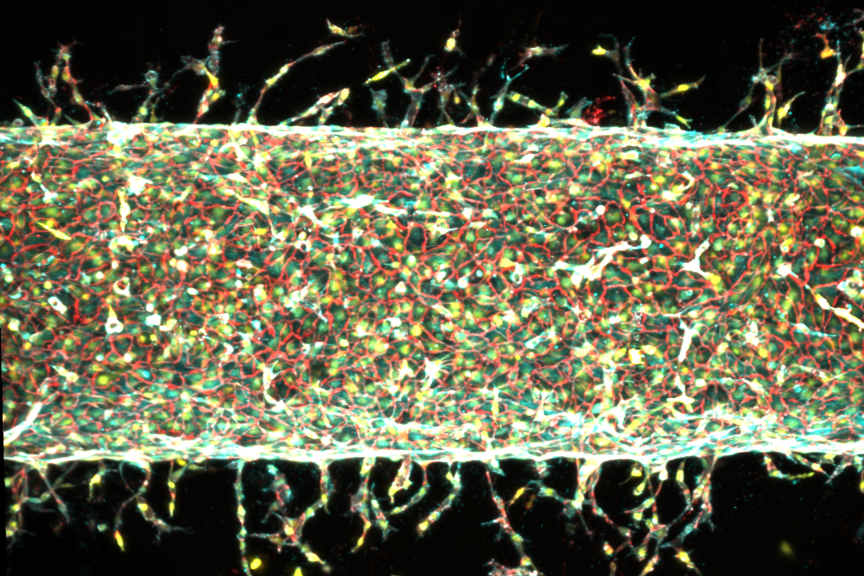
The results of these experiments were compelling and offered clear evidence of mechanical control over angiogenesis. When the main artery within the gel was simply left undisturbed, it did sprout some new vessels, but these appeared in random locations and without any discernible pattern. However, when the artery was subjected to the controlled jostling, the difference was stark: significantly more vessels sprouted, indicating that mechanical stimulation actively enhances the angiogenic process.
The team further refined their experiments to understand how specific mechanical parameters influenced vessel morphology. They found that stretching the gel by 5 percent of its total width resulted in a proliferation of many new vessels emerging from the main artery. This level of stretch appeared to optimize the sheer number of capillaries formed. In contrast, when the stretch was increased to 15 percent, fewer vessels sprouted overall, but those that did grow were noticeably longer. This suggests a nuanced control mechanism where different degrees of mechanical strain can tune the angiogenic response, influencing both vessel density and length.
Perhaps the most significant finding regarding control was the ability to direct the growth trajectory of these new vessels. When the researchers changed the direction of the mechanical stretching, the newly sprouting vessels responded in kind, taking turns and meticulously following the imposed pattern of stimulation. This unprecedented level of directional control is a critical advancement, as it means scientists could potentially "program" the intricate branching patterns required for complex engineered tissues. "We’re finding that moving is good, which is always the takeaway of everything we do in our lab," Raman notes, summarizing the consistent theme across her lab’s research. "Mechanical forces play an important role in our bodies. That means that if you want to grow more or less vessels, or shorter or longer vessels, or vessels in certain directions, we now know how to do that." This statement underscores the practical, actionable knowledge derived from their research.
Unraveling the Mechanism: The Role of Piezo1
Beyond simply observing the phenomenon, the researchers delved deeper to understand the underlying biological mechanisms driving this mechanically induced angiogenesis. Their investigation led them to a specific gene and its associated protein: Piezo1.
The connection to Piezo1 arose from Raman’s attendance at a talk by molecular biologist Ardem Patapoutian. In 2021, Patapoutian was awarded the Nobel Prize in Physiology or Medicine for his groundbreaking discovery of mechanosensitive ion channels in cell membranes, specifically PIEZO1 and PIEZO2. These channels act as molecular "gatekeepers" that open and close in response to mechanical pressure, controlling the flow of ions into and out of cells. This ion flux can trigger various cellular responses.
After Patapoutian’s presentation, Raman shared her group’s experimental results, which clearly demonstrated a link between blood vessel growth and mechanical stimulation. Patapoutian, recognizing the parallel with his own work, proposed a compelling hypothesis: the mechanical exercise applied to the central artery might be stimulating PIEZO1 ion channels in the endothelial cells, thereby triggering the growth of new blood vessels.
To rigorously test this hypothesis, Raman’s team embarked on a gene-editing experiment. Their strategy was to "knock down" the PIEZO1 gene, reducing its activity. If the PIEZO1 channel was indeed the critical mediator, then suppressing its function should lead to a reduction in mechanically induced blood vessel growth. The team repeated their experiments, this time using endothelial cells that had been genetically engineered to express less of the PIEZO1 gene. The results provided strong validation for Patapoutian’s hypothesis: even with continuous mechanical exercise of the central artery, significantly fewer new blood vessels sprouted from the genetically modified cells. This confirmed that PIEZO1 ion channels are crucial for transducing mechanical signals into angiogenic responses, providing a clear molecular pathway for the observed phenomenon.
Broader Implications: A New Era for Engineered Tissues and Beyond
The implications of this discovery extend far beyond the laboratory, promising to usher in a new era for regenerative medicine, artificial organ development, and disease modeling. The ability to precisely control the growth and patterning of blood vessels using physical cues represents a paradigm shift from the more unpredictable chemical-based approaches.
- Regenerative Medicine: For patients suffering from tissue damage due to injury, disease, or aging, this technology could be transformative. Imagine creating custom-engineered skin grafts with built-in vascular networks for severe burn victims, or regenerating muscle tissue with a pre-formed blood supply to restore function after debilitating injuries. Chronic wounds, which often fail to heal due to inadequate blood flow, could potentially benefit from vascularized tissue implants.
- Artificial Organ Development: One of the most significant bottlenecks in developing fully functional artificial organs for transplantation—such as lab-grown kidneys, livers, or hearts—is the lack of an integrated vascular system. Without a dense network of capillaries to sustain them, these complex organs cannot survive or function once implanted. This new method offers a viable pathway to engineer these essential vascular networks, potentially bringing lab-grown organs closer to clinical reality and addressing the critical shortage of donor organs.
- Disease Modeling: The creation of more physiologically relevant in vitro disease models is crucial for drug discovery and understanding disease progression. With the ability to grow organized vascular networks, researchers can develop microphysiological systems (organs-on-a-chip) that more accurately mimic the human body’s environment. This could lead to better testing of new drugs for efficacy and toxicity, particularly in conditions involving vascular dysfunction, such as atherosclerosis, diabetes-related microvascular complications, or even tumor angiogenesis.
- Biomaterials and Implants: The insights gained from this research could also inform the design of next-generation biocompatible implants. By incorporating mechanosensitive elements or designing implant surfaces that encourage specific mechanical interactions with surrounding cells, it might be possible to stimulate the body’s own vascular system to integrate more effectively with prosthetic devices.
- Fundamental Biological Understanding: Beyond its immediate applications, this research deepens our understanding of mechanobiology—how physical forces influence cellular behavior and tissue development. It highlights the profound and often underestimated role of mechanical cues in orchestrating complex biological processes like angiogenesis.
Looking ahead, the team is already planning the next phases of their research. Co-author Jessica Shah states, "We are now investigating how precisely patterning blood vessel growth can help improve muscle function." This suggests a direct translational path, combining their earlier work on artificial muscles with this new capability in vascular engineering. The collaborative nature of this research, involving co-authors from both MIT (Sina Kheiri, Jessica Shah, Shashaank Venkatesh, and Roger Kamm) and Harvard University (Peiyuan Chai and Ryan Flynn), underscores the interdisciplinary effort required for such complex biological engineering challenges.
This pioneering work, supported in part by significant funding from the U.S. Department of War Army Research Office Early Career Program, PECASE Grant, and a Department of War DURIP Program Grant, positions mechanical stimulation as a powerful, precise, and scalable tool for tissue engineering. The ability to program blood vessel growth with physical cues offers a robust pathway for the reproducible fabrication of engineered tissues that could ultimately be implanted in the body, restoring function after debilitating disease or injury and ushering in a new era of regenerative medicine.