Cambridge, MA / Baltimore, MD – A significant advancement in the battle against one of the most lethal cancers, ovarian cancer, has emerged from a collaborative effort between researchers at MIT and Johns Hopkins University. A new handheld microfluidic device has been developed, capable of gently collecting living cells from specific tissue locations, offering unprecedented potential for earlier and more effective cancer detection, as well as the development of personalized treatments. This innovation directly addresses the critical challenge of ovarian cancer’s high mortality rate, which is largely attributed to its late-stage diagnosis.
The devastating impact of ovarian cancer stems from a cruel paradox: when detected in its earliest stages, the five-year survival rate for patients can soar to over 90 percent. However, this promising outlook plummets dramatically, often falling below 50 percent, once the disease progresses to advanced stages (III or IV). This stark disparity underscores the urgent need for improved diagnostic tools, a need that the new device, detailed in a study published in the journal Device, aims to fulfill.
The Elusive Enemy: Why Ovarian Cancer Remains a Silent Killer
Ovarian cancer, often dubbed the "silent killer," is the fifth leading cause of cancer-related death among women, accounting for more deaths than any other cancer of the female reproductive system. Globally, it affects hundreds of thousands of women each year, with incidence rates varying by region and ethnicity. In the United States, for instance, approximately 20,000 women are diagnosed annually, and over 13,000 succumb to the disease. The primary reason for this grim statistic is the absence of clear, early symptoms and the lack of an effective, widespread screening method for the general population. Symptoms such as bloating, pelvic or abdominal pain, difficulty eating, or feeling full quickly are often vague and can be easily mistaken for less serious conditions, leading to significant diagnostic delays.
Current screening methods, including transvaginal ultrasound (TVUS) and the CA-125 blood test, have proven insufficient for early detection in asymptomatic women. TVUS can identify masses but cannot definitively distinguish between benign and malignant growths, often leading to unnecessary surgeries. CA-125, a protein biomarker, can be elevated in various non-cancerous conditions and is not always elevated in early-stage ovarian cancer, making it an unreliable standalone screening tool. This diagnostic gap has long been a major hurdle for oncologists and patients alike, emphasizing the critical importance of novel approaches to detect the disease when it is most treatable.
A Paradigm Shift: Tracing Ovarian Cancer to Its Origins
A pivotal breakthrough occurred approximately two decades ago when researchers made a groundbreaking discovery: many cases of high-grade serous ovarian cancer (HGSOC), the most common and aggressive subtype of ovarian cancer, do not originate in the ovary itself, but rather in the fimbriae (finger-like projections) at the ends of the fallopian tubes. This understanding represented a significant paradigm shift, challenging long-held beliefs about the disease’s genesis. These precursor lesions, known as Serous Tubal Intraepithelial Carcinoma (STICs), are often microscopic and exceedingly difficult to detect or sample using conventional methods.
This revelation has already had tangible impacts on prevention strategies, particularly for women at high genetic risk, such as those with BRCA1 or BRCA2 mutations. For these individuals, who face a significantly elevated lifetime risk of ovarian cancer, prophylactic salpingectomy (surgical removal of the fallopian tubes, often while preserving the ovaries to maintain hormone production) after childbearing years has emerged as a crucial risk-reduction option. However, for the broader population and for identifying nascent disease in symptomatic women, the challenge of detecting these microscopic, pre-cancerous lesions in situ has persisted.
The Innovation: A Gentle Touch for Precise Cellular Insights
It is against this backdrop that the new handheld device developed by MIT Professor Kripa Varanasi’s research group, in collaboration with colleagues at MIT and Johns Hopkins University, promises to revolutionize early detection. The device is designed to gently collect living cells from highly specific, microscopic locations within tissues, a capability that traditional biopsy methods often lack or compromise.
"We wanted to collect living cells from specific regions of the fallopian tube while leaving the surrounding tissue intact," explains Professor Varanasi, the senior author of the study and the Maher A. Elmasri Professor of Mechanical Engineering. "Once we have these living cells, there are many things we can do with them. We can use them for diagnostics, grow them into organoids, and build living models of disease. Ultimately, this could allow us to test how an individual patient’s cells respond to different treatments and help us develop more personalized medicines."
The device operates on a sophisticated microfluidic principle. It features a small microfluidic channel that is gently held against the tissue. A syringe then drives fluid through this channel, applying a precisely controlled shear force parallel to the tissue surface. This delicate, targeted force is sufficient to detach living cells from tiny sections of tissue without causing significant damage to the surrounding cellular architecture. This method stands in stark contrast to conventional biopsy techniques, which often involve excising larger tissue samples or using abrasive brushes that can damage cells and compromise their viability.
A Collaborative Endeavor: Bridging Engineering and Oncology
The development of this innovative technology is a testament to the power of interdisciplinary collaboration. The project brought together expertise from diverse fields: mechanical engineering, biological engineering, materials science, and gynecologic oncology. Professor Angela Belcher, MIT’s James Mason Crafts Professor and a professor of biological engineering and materials science and engineering, played a crucial role in connecting Professor Varanasi with Dr. Rebecca Stone, the Stoddard and O’Neil Professor in Gynecologic Oncology at Johns Hopkins University School of Medicine.
This powerful trio, along with their respective teams, secured funding from Break Through Cancer, a foundation dedicated to fostering interdisciplinary research to tackle challenging cancer problems. Their shared vision centered on developing novel approaches for the early detection of ovarian cancer, with this new cell-collection technology forming a cornerstone of their broader effort.
Dr. Stone, a long-time advocate for preventive salpingectomy in high-risk women, emphasizes the clinical significance of such a device. "This is exactly the kind of problem that benefits from bringing clinicians and engineers together," Stone notes. "We understand the clinical need, while the MIT team brings a very different perspective from fluid mechanics and engineering. That combination allowed us to approach the problem in a new way."
Overcoming the Limitations of Traditional Pathology
The current standard practice for examining surgically removed fallopian tubes involves placing the tissue in a chemical preservative, then meticulously cutting it into sections for microscopic analysis. While this method preserves tissue structure for pathological examination, it renders the cells non-viable, meaning they cannot be grown in culture or used for functional studies. This process is not only time-consuming but also inherently destructive to the cellular integrity required for advanced research.
"It’s very time-consuming and destructive to the cells," Varanasi observes. "We wanted to bring new capabilities to pathology, so we can not only see what these cells look like, but also collect them alive and study how they behave." The inspiration for the fluid-based approach came to the MIT researchers while observing surgeons in operating rooms at Johns Hopkins, noting how easily some cells detached during routine procedures. They realized that a controlled fluid flow could leverage this natural cellular property more effectively than a scalpel or brush.
The researchers developed a 3D-printed microfluidic device that forms a vacuum seal with the tissue, confining the liquid flow to a precise, small region. As Bert Vandereydt, a co-first author and MIT postdoc, explains, "We came up with this device where one syringe creates a vacuum that holds it against the tissue, and a second syringe pushes liquid through it. The vacuum creates a seal, so nothing leaks, and then we locally apply what is basically a microfluidic chip on the tissue that gently shears the cells off."
Proof of Concept and Clinical Application
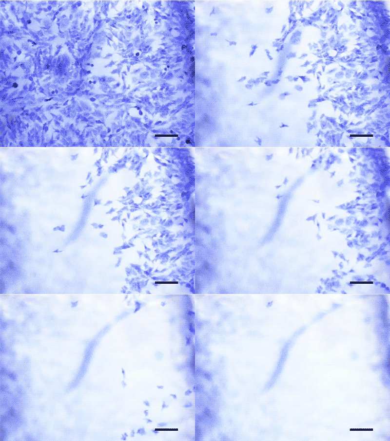
To validate their innovative system, the researchers rigorously tested the device, demonstrating its ability to tune the applied shear stress and comparing its performance against conventional cell collection workflows. Critically, they found that cells collected using their microfluidic technique remained highly viable and exhibited significantly better growth rates in culture compared to cells obtained via traditional methods.
The ultimate test involved applying the device to fresh human fallopian tube samples, a process that required close coordination with their Johns Hopkins collaborators, often involving urgent, late-night notifications for sample shipments. "The samples could come at any time. Sometimes, we’d get an email from our collaborators at 11 p.m. saying ‘There are two fallopian tubes coming tomorrow,’" Vandereydt recounts. Despite the logistical challenges, the team successfully collected living cells from these samples and, crucially, developed them into organoids – miniature, self-organizing 3D cultures that mimic the structure and function of actual organs. This capability is paramount for disease modeling, drug screening, and ultimately, developing personalized treatments tailored to an individual patient’s unique cellular response.
Professor Belcher highlights the potential synergy with other diagnostic technologies: "We are developing optical approaches to identify suspicious regions of tissue, and this technology could allow us to collect living cells from exactly those locations. Being able to first see where the disease may be emerging and then collect those cells for further study could be very powerful."
Beyond Ovarian Cancer: A Versatile Diagnostic Platform
One of the most promising aspects of this new technology is its versatility. The researchers demonstrated that the device is "agnostic to the disease," meaning it can be tuned to collect various cell types by adjusting the level of shear stress. As Vandereydt explains, "There are very loosely adherent prostate cancer cells that detach at 1 pascal [of stress], but if you look at bone cancer cells, only a few cells detach under as high as 5 pascals of stress." This adaptability suggests that the microfluidic cell collection system could have broad applications across a spectrum of cancers and other diseases where cellular analysis is critical.
Implications for Personalized Medicine and Future Diagnostics
The ability to obtain viable, patient-specific cells from precise locations opens up transformative avenues in cancer care.
- Personalized Treatment Regimens: By cultivating organoids from a patient’s own living cancer cells, clinicians could test various chemotherapy drugs or targeted therapies in vitro, effectively creating a "drug-response profile" for that individual. This could eliminate trial-and-error approaches, reduce treatment side effects, and significantly improve therapeutic outcomes, ushering in a new era of truly personalized oncology.
- Enhanced Early Detection: While initial applications are envisioned for ex vivo tissue (tissue already removed from the body, simplifying regulatory approval), the long-term goal is to integrate the device into in vivo diagnostic procedures. Imagine a future where a miniature version of this device could be deployed via endoscopy or laparoscopy to swab suspicious areas within the body, offering a non-invasive or minimally invasive way to detect pre-cancerous lesions or early-stage cancers before they become widespread.
- Deeper Understanding of Carcinogenesis: The ability to study living cells from the earliest stages of disease development provides an unprecedented window into the fundamental biology of cancer. Researchers can monitor cellular changes, identify early biomarkers, and unravel the complex molecular pathways that drive tumor progression, leading to new therapeutic targets and preventive strategies.
- Integration into Pathology: Dr. Stone envisions integrating this technology into routine histopathology workflows. Instead of solely relying on static, preserved tissue sections, pathologists could gain dynamic insights into cellular behavior and viability, enhancing their diagnostic capabilities and fostering a more comprehensive understanding of disease.
- Reduced Healthcare Burden: Earlier and more precise diagnosis could lead to less extensive and less costly treatments, significantly reducing the financial and emotional burden on patients and healthcare systems alike. The potential to prevent advanced disease progression could save countless lives and improve quality of life for cancer patients.
Challenges and the Road Ahead
While the promise of this technology is immense, the path from device to widespread diagnostic tool will involve several critical steps. Regulatory approval, particularly for in vivo applications, will require rigorous clinical trials to demonstrate safety and efficacy in human subjects. Further miniaturization and integration with existing endoscopic or surgical instruments will be necessary for widespread clinical adoption. Additionally, considerations around cost-effectiveness and accessibility will be vital to ensure that this groundbreaking technology can benefit all who need it.
The researchers, driven by a profound sense of purpose, remain optimistic. Professor Varanasi concludes, "If this work can ultimately help women by enabling earlier detection of ovarian cancer, I would find that incredibly fulfilling. That is really what motivates us — taking the science and engineering we develop in the lab and using it to make a difference in people’s lives."
This project, supported by the Break Through Cancer Foundation, stands as a beacon of hope, illustrating how collaborative, interdisciplinary research can push the boundaries of medical science and pave the way for a future where cancers like ovarian cancer are caught early, treated effectively, and ultimately, conquered. The journey has just begun, but the initial steps suggest a future where the silent killer might finally be heard, and stopped, in its tracks.