Engineers at the Massachusetts Institute of Technology (MIT) have achieved a significant breakthrough in biohybrid robotics, demonstrating that even a single, ultrathin layer of muscle cells can generate enough force to power a swimming robot through water. This innovation, detailed in a paper published today in the journal Advanced Functional Materials, presents a novel design for a muscle-powered aquabot, marking a pivotal step towards developing soft, self-healing robots capable of navigating complex and delicate environments.
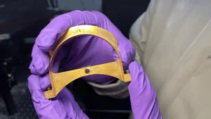
At the core of this groundbreaking device is a flexible gel film, approximately the length and width of a standard stick of gum, which serves as the robot’s "skeleton." This film is ingeniously designed with two distinct halves, forming the bot’s "fins." Each fin is meticulously covered with a layer of live muscle cells, astonishingly thin – far less than the diameter of a single human hair. These specialized cells are not just any muscle tissue; they have been genetically engineered to respond and twitch specifically when exposed to light. The mechanism of locomotion is elegantly simple yet profoundly effective: by precisely shining a light on one fin, the embedded muscles contract, causing the fin to flap with sufficient force to propel the robot through water. The direction and speed of the robot can be finely tuned by alternating the light flashes between the two fins at various intervals, offering a degree of controlled movement previously unseen in such minimalist biohybrid designs.
During initial trials, the paper-thin robot showcased its capabilities by successfully swimming and swiveling through a basic watery maze. While its top speed of approximately four times its body length per minute might seem modest when compared to the blistering pace of Olympic swimmers, who can cover up to 65 body lengths per minute, it holds its own against more leisurely marine life. For instance, the robot’s speed is comparable to that of a cow shark, known for its unhurried exploration of ocean depths. Ritu Raman, an associate professor of mechanical engineering at MIT and a lead author of the study, emphasized the inherent strength of the design. "It takes a lot of force to move through water versus air," Raman noted. "The robot’s quite strong, given its size." This statement underscores the efficiency and power generated by the remarkably thin muscle layer.
A New Paradigm in Biohybrid Robotics
This new robot represents a significant advancement, being the first example of a very thin, two-dimensional, muscle-powered robot capable of sustained locomotion. Traditional biohybrid robots, including earlier models developed by Raman’s group and others, have typically relied on bulky, three-dimensional chunks of lab-grown skeletal muscle, necessitating millions of cells for their fabrication. Such designs often present challenges in terms of cost, manufacturing complexity, and energy efficiency. Raman highlighted the advantages of their thinner, less bulky design: "Thinner, less bulky designs such as the team’s new bot could be cheaper to build and could move more efficiently." This cost-effectiveness and enhanced efficiency are crucial for the broader adoption and scalability of biohybrid technologies.
The long-term vision for these biohybrid robots extends beyond simple aquatic navigation. Raman articulated the profound potential: "We believe that biohybrid robots powered by living muscle could one day perform delicate jobs like exploring environments too fragile or unpredictable for conventional hardware, because living tissue is soft, responsive to its surroundings, and can heal itself." This vision positions biohybrid robots as ideal candidates for tasks requiring innate compliance, adaptability, and even self-repair capabilities, attributes inherently lacking in rigid, purely mechanical systems. Such applications could include targeted drug delivery within the human body, environmental monitoring in sensitive ecosystems, or intricate inspections in hazardous industrial settings. The study’s co-authors from MIT include first author Maheera Bawa, Arielle Berman, Laura Schwendeman, Ferdows Afghah, and Seanbiron Johnson, whose collective efforts were instrumental in bringing this innovation to fruition.
Tracing the Path: From Static Actuation to Dynamic Locomotion
The development of this swimming robot is built upon a foundation of prior research from Raman’s group, specifically their work from the previous year on an iris-inspired disk of artificial muscle tissue. In that earlier project, the team developed a gel disk stamped with a pattern of concentric and radial grooves. Live muscle cells were then deposited onto the gel’s surface, where they grew into a thin layer, aligning themselves along these intricate grooves. When stimulated with light, these cells moved in coordinated patterns, causing the disk to stretch and squeeze in a manner reminiscent of how a human iris dilates and constricts the eye’s pupil.
That pioneering work was groundbreaking in demonstrating that muscle cells could be grown in extremely thin layers and configured into complex patterns, allowing for controllable, multi-directional movement upon stimulation. "People hadn’t seen this muscle architecture engineered from scratch before," Raman explained, emphasizing the novelty of their approach. "And the cells were moving in multiple directions." However, a significant limitation of this initial design was the scale of movement; the cells only moved approximately 100 microns. While impressive for cellular actuation, this was deemed insufficient from a robotics perspective to achieve macroscopic locomotion. "From a robotics perspective, their movements were tiny," Raman conceded. This recognition spurred the team’s subsequent efforts to significantly amplify muscle movements, aiming to generate enough force to power a self-propelling robot. The key to achieving this, they hypothesized and subsequently proved, lay in meticulously optimizing the "skeleton" – the underlying gel structure – upon which the muscle cells were cultivated.
The Art of Optimization: Tuning the Biohybrid Skeleton
To transition from microscopic actuation to robust macroscopic movement, the MIT team embarked on a systematic optimization of the underlying gel structure, which they aptly refer to as the "skeleton." Their strategy focused on tuning three critical properties of the gel: its chemical composition, its mechanical stiffness, and the precise size and shape of the grooves stamped into its surface. These parameters are well-known in tissue engineering as crucial "knobs" that can be adjusted to influence cell growth, alignment, and force generation. "For engineering any type of tissue, it’s known that these are knobs you can tune," Raman stated. "And we wanted to optimize all these parameters to support live muscle cells."
The first area of investigation involved the groove geometry. The researchers experimented with various groove types, contrasting, for instance, a skinny square trough with a more elongated, curved valley. Their findings revealed that muscle cells exhibited superior alignment and organization when deposited onto gels featuring grooves that were more square in cross-section than curved. This enhanced alignment is crucial because more aligned cells tend to fuse into cohesive fibers, forming a stronger, more coordinated muscle tissue capable of generating greater collective force. Thus, square grooves emerged as the optimal design choice.
Next, the team addressed the gel composition. Their previous iris-inspired design had utilized fibrin, an ultrasoft gel. While biocompatible, fibrin proved to be problematic as it could quickly shrivel in response to the contractile forces generated by the muscles, thereby limiting the overall movement and stability. To overcome this, they turned to gelatin methacrylate (GelMA), a material widely employed in tissue engineering due to its excellent biocompatibility and tunable mechanical properties. By formulating different recipes of GelMA, the team was able to create skeletons with varying degrees of stiffness. They meticulously observed how muscle cells grew and performed on these diverse GelMA surfaces. The results were clear: cells exhibited better alignment and produced significantly more force when grown on stiffer gels, indicating that a robust foundation was essential for maximizing muscle performance.
The gel thickness was another critical parameter. The team experimented with different thicknesses, ultimately identifying that a half-millimeter-thin film of GelMA provided the ideal balance. This thickness offered sufficient structural support for a single layer of muscle cells while remaining light enough to prevent the cells from peeling away from the gel surface during contraction, ensuring efficient force transmission.
Finally, to further enhance the muscle’s capabilities, the team incorporated a unique "exercising" regimen. Similar to how physical training strengthens human muscles, the researchers subjected the biohybrid muscles to a training routine involving programmed sequences of flashing lights. This regular stimulation helped to strengthen the muscle cells, increasing their contractile force and overall robustness.
With these optimized parameters – pumped-up cells on a meticulously tuned GelMA skeleton featuring square-bottomed grooves – the team proceeded to design the thin, two-finned robot. The muscle cells, aligned by the grooves, fused into strong, cohesive fibers, forming a powerful, integrated muscle tissue on each fin. "You can think of the robot as having two independent muscles," Raman explained, highlighting the modular control. "If we shine a light on just one, only that muscle moves. If shining on both, they both flap." This independent actuation is what enables precise steering and speed control.
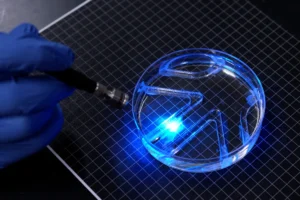
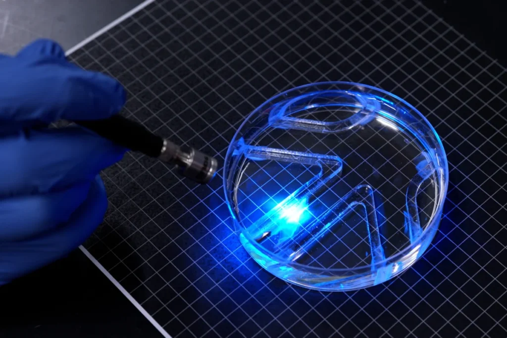
To demonstrate the robot’s navigational abilities, the researchers submerged it in a large petri dish filled with water. They then manually maneuvered a light source over the bot, which faithfully followed the light, flapping its fins in response to navigate successfully through a simple maze placed within the dish. This demonstration underscored the effective integration of the biological and engineered components, showcasing a tangible step forward in controllable biohybrid locomotion.
Broader Implications and the Future of Biohybrid Robotics
This breakthrough by MIT engineers holds profound implications for the nascent but rapidly evolving field of biohybrid robotics. The ability to create functional, locomoting robots from single-layer muscle tissue addresses several critical challenges that have historically hampered the development of such systems.
Firstly, the reduced cellular requirement significantly lowers the cost and complexity of fabrication. Current biohybrid robots often demand vast quantities of cells and intricate 3D bioprinting techniques, making them expensive and difficult to scale. By demonstrating effective locomotion with minimal biological material, the MIT team paves the way for more accessible and widely applicable biohybrid systems.
Secondly, the inherent softness and responsiveness of living tissue offer advantages that traditional rigid robots cannot match. Biohybrid robots are intrinsically compliant, allowing them to interact safely with delicate objects or navigate constricted, irregular spaces without causing damage. Their responsiveness to environmental cues, particularly through engineered cells, opens possibilities for adaptive behaviors that are difficult to program into conventional hardware. Furthermore, the self-healing capacity of living tissue is a revolutionary attribute. Unlike mechanical robots that require repairs or component replacement after damage, biohybrid systems could potentially mend themselves, extending their operational lifespan and reducing maintenance costs, especially in remote or inaccessible environments.
The development also pushes the boundaries of soft robotics. While many soft robots rely on pneumatic or hydraulic actuation, biohybrid systems introduce a new paradigm of biological power, potentially leading to more energy-efficient and autonomously powered soft machines. This could revolutionize areas like medical micro-robotics, where devices need to navigate complex biological systems without causing trauma, or in environmental exploration, where biodegradable and self-sustaining robots could monitor ecosystems without leaving behind persistent waste.
However, the path forward is not without its challenges. Scaling up these devices to larger sizes or for more complex tasks will require advancements in nutrient delivery systems to sustain the living cells over extended periods. Ensuring the long-term viability and functionality of the muscle tissue outside a controlled laboratory environment remains a key hurdle. Integrating sophisticated sensing capabilities and more complex control algorithms to enable autonomous decision-making are also critical next steps. Ethical considerations surrounding the use of living tissues in robotic systems, while nascent for simple muscle-powered bots, will also become increasingly relevant as these technologies advance towards more complex "living machines."
In conclusion, the MIT team’s achievement in creating a thin, muscle-powered swimming robot represents a pivotal moment in biohybrid robotics. By optimizing the bio-interface between living muscle cells and an engineered skeleton, they have unlocked a new level of efficiency and control in biologically actuated systems. This innovation not only expands the potential applications for soft, adaptive robots but also brings humanity closer to a future where machines can mimic the intricate functionalities and resilience of living organisms, opening doors to exploration, medical intervention, and environmental stewardship in ways previously unimaginable.