3D bioprinting, a transformative technology that involves fabricating living tissues by extruding cells suspended within hydrogel-based “bio-inks,” is rapidly becoming a cornerstone of bioengineering. Its applications span from creating intricate tissue models for disease research and drug development to the ambitious goal of replacing damaged or diseased tissues within the human body. However, the precision and reproducibility of these printed tissues are often hampered by a persistent challenge: gravity. The inherent density difference between cells and their surrounding hydrogel matrix causes cells to settle within the printer’s extrusion syringe, leading to a cascade of issues that compromise print quality and tissue consistency.
This phenomenon, known as cell sedimentation, is particularly problematic during the extended printing sessions required for larger or more complex tissue constructs. As cells drift to the bottom of the syringe, they can lead to nozzle blockages, resulting in uneven cell distribution throughout the printed material. This unevenness directly impacts the biological function and structural integrity of the resulting tissue, creating significant inconsistencies between different printed samples.
Ritu Raman, the Eugene Bell Career Development Professor of Tissue Engineering and an assistant professor of mechanical engineering at MIT, has been at the forefront of addressing this critical limitation. “This cell settling, which becomes worse during the long print sessions required to print large tissues, leads to clogged nozzles, uneven cell distribution, and inconsistencies between printed tissues,” Raman explained. Current mitigation strategies, such as manual stirring of bio-inks prior to printing or the use of passive mixing mechanisms, prove insufficient. These methods can only maintain homogeneity for a limited time, failing to counteract the relentless force of gravity once the printing process is underway.
A groundbreaking study, published on February 2, 2024, in the journal Device, introduces a novel solution developed by Raman’s team. This innovative approach actively combats cell sedimentation within bio-inks during the printing process, paving the way for more reliable and biologically consistent 3D bioprinted tissues. The research, titled "Advancing Bioink Homogeneity in Extrusion 3D Bioprinting with Active In Situ Magnetic Mixing," details the development and validation of a system designed to ensure uniform cell distribution from the initial loading of the bio-ink to the final layer of the printed construct.
The Challenge of Gravity in Bioprinting
The fundamental principle of 3D bioprinting relies on precise control over the physical and biological properties of the bio-ink. Ferdows Afghah, a postdoctoral researcher in mechanical engineering at MIT and the lead author of the study, emphasized this point: “Precise control over the bioink’s physical and biological properties is essential for recreating the structure and function of native tissues.” The weight disparity between cellular components and the hydrogel carrier is a persistent hurdle. Typical bio-inks are composed of biocompatible polymers, such as gelatin, alginate, or hyaluronic acid, which form a gel-like matrix to provide structural support and a conducive environment for cell survival and proliferation. While these hydrogels are crucial for mimicking the extracellular matrix of natural tissues, they are generally less dense than the cells they encapsulate.
Over time, and exacerbated by the forces involved in extrusion, this density difference causes the heavier cells to migrate downwards within the syringe. This sedimentation can significantly alter the cell-to-matrix ratio in different parts of the printed construct. For applications in disease modeling, for instance, an uneven distribution of cells could lead to inaccurate representations of pathological conditions, potentially skewing research findings. In drug screening, inconsistent cell density might result in variable cellular responses to therapeutic agents, making it difficult to ascertain the true efficacy or toxicity of a drug.
Introducing MagMix: An Active Mixing Solution
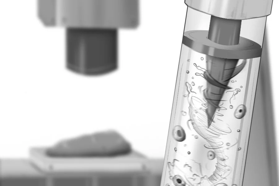
To overcome the limitations of passive mixing, Raman’s team developed "MagMix," a magnetically actuated mixer. This compact system consists of two primary components: a small magnetic propeller designed to fit inside the bio-ink syringe, and an external permanent magnet attached to a motor. The motor-driven magnet is positioned to move vertically along the exterior of the syringe, precisely controlling the rotation and movement of the internal propeller. This dynamic interaction ensures that the bio-ink is continuously and uniformly agitated throughout the printing process.
The MagMix system is engineered for seamless integration with existing 3D bioprinting platforms. It can be mounted onto standard bioprinters without requiring any modifications to the printer’s hardware or the bio-ink formulation itself. This adaptability makes it a broadly accessible solution for a wide range of research laboratories and industrial settings. The design prioritizes minimal interference with the printer’s normal operation, ensuring that the bioprinting process remains as straightforward as possible.
The development process involved a meticulous combination of computational modeling and experimental validation. The team utilized computer simulations to optimize the geometry of the magnetic propeller and determine the ideal mixing speeds required to achieve effective homogenization without causing undue stress on the cells. These simulations allowed for a data-driven approach to design, ensuring that the final physical device would perform optimally. Following the simulation phase, the team rigorously tested the MagMix system experimentally to confirm its efficacy in real-world bioprinting scenarios.
Experimental Validation and Performance
The experimental results demonstrated the remarkable effectiveness of MagMix. “Across multiple bioink types, MagMix prevented cell settling for more than 45 minutes of continuous printing, reducing clogging and preserving high cell viability,” reported Raman. This sustained mixing capability is a significant leap forward, addressing the challenges posed by long printing sessions. The system was shown to effectively counteract gravitational settling, maintaining a uniform distribution of cells within the bio-ink.
Crucially, the researchers were able to fine-tune the mixing speeds to achieve a delicate balance. They demonstrated that the system could be adjusted to provide effective homogenization for various bio-ink formulations while inducing minimal mechanical stress on the sensitive cellular components. High cell viability is paramount for the success of bioprinting applications, as damaged or dead cells cannot contribute to tissue development or function.
As a proof-of-concept, the team successfully utilized MagMix to 3D print cells that, over several days, matured into functional muscle tissues. This demonstration highlights the system’s ability to support the biological processes necessary for tissue development post-printing, a critical factor for regenerative medicine applications. The consistent cell distribution enabled by MagMix is directly correlated with the enhanced biological function and structural integrity of the printed tissues.
Implications for Disease Modeling, Drug Screening, and Regenerative Medicine
The ability to fabricate high-quality tissues with consistent biological function has profound implications across several critical areas of biomedical research and development.
-
Disease Modeling: By creating more accurate and reproducible 3D tissue models, researchers can gain deeper insights into the mechanisms of human diseases. These models can mimic the complex cellular microenvironments found in native tissues, allowing for the study of disease progression, the identification of novel therapeutic targets, and the testing of interventions in a more physiologically relevant context than traditional 2D cell cultures.
-
Drug Screening and Toxicology: The development of robust tissue models that closely resemble human organs or tissues is a key objective for the pharmaceutical industry. Such models can significantly accelerate the process of drug discovery and development by providing a platform for high-throughput screening of potential drug candidates. Moreover, these models can be used to assess the safety and efficacy of new therapeutic drugs, potentially reducing the reliance on animal testing. This aligns with the growing interest from regulatory bodies like the U.S. Food and Drug Administration (FDA) in developing "New Approach Methodologies" (NAMs) that are faster, less expensive, and more informative for evaluating drug safety and efficacy.
-
Regenerative Medicine: The ultimate goal of much of this research is the development of functional 3D-printed tissues that can be implanted into patients to replace diseased or injured tissues. Raman elaborated on this ambitious vision: “Eventually, we are working towards regenerative medicine applications such as replacing diseased or injured tissues in our bodies with 3D printed tissues that can help restore healthy function.” The consistency and reliability afforded by MagMix are essential for translating this vision into clinical reality, ensuring that implanted tissues perform as intended and integrate effectively with the host body.
The Role of the Safety, Health, and Environmental Discovery Lab (SHED)
The development of MagMix was significantly supported by the Safety, Health, and Environmental Discovery Lab (SHED) at MIT. This interdisciplinary hub provides essential infrastructure and expertise to accelerate the translation of biofabrication innovations from laboratory-scale demonstrations to scalable and reproducible applications.
Tolga Durak, the founding director of the SHED, highlighted the lab’s mission: “At the SHED, we focus on accelerating the translation of innovative methods into practical tools that researchers can reliably adopt.” He views MagMix as a prime example of how interdisciplinary support and robust technical infrastructure can drive biofabrication technologies toward real-world impact. The SHED’s involvement underscores a broader commitment to strengthening technology pathways that enhance reproducibility and accessibility in both engineering and the life sciences. This includes providing equitable access to advanced equipment and fostering cross-disciplinary collaboration, which are crucial for overcoming the complex challenges inherent in translating novel research into practical applications.
Durak further emphasized the importance of integrated labs like SHED in building sustainable capacity as the field of bioprinting advances toward larger-scale and more standardized systems. “Our goal is not only to enable discovery, but to ensure that new technologies can be reliably adopted and sustained over time,” he stated. This focus on long-term viability and widespread adoption is critical for the field’s continued progress.
Beyond Medical Applications: Biohybrid Robots
The potential applications of MagMix extend beyond human health. The research team is also exploring the use of engineered tissues, such as printed muscles, to power innovative "biohybrid" robots. These robots, which combine biological components with artificial systems, could offer new possibilities for creating safer, more efficient, and potentially more sustainable robotic technologies. For instance, bio-actuated systems might be employed in applications where traditional motors are impractical or undesirable, such as in delicate environmental monitoring or biomedical devices.
Future Outlook and Conclusion
The development of MagMix represents a significant advancement in the field of 3D bioprinting. By effectively addressing the fundamental challenge of cell sedimentation, this technology promises to improve the reliability and scalability of printed tissues. The potential impacts on both the scientific understanding of tissue engineering and the practical applications for human health are substantial.
The researchers are optimistic that their work will pave the way for more consistent and functional engineered tissues. This, in turn, will accelerate progress in critical areas such as disease modeling, drug discovery, and the ultimate realization of regenerative medicine therapies. The publication of their findings in the journal Device marks a key milestone, making this innovative technology and its supporting data accessible to the broader scientific community. The ongoing collaboration with facilities like the SHED further ensures that such advancements are not just theoretical breakthroughs but are poised for practical implementation and widespread adoption, driving forward the future of biofabrication.