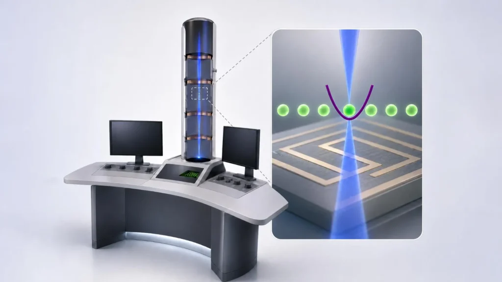
The landscape of high-resolution imaging is currently undergoing a fundamental shift as researchers in Austria bridge the gap between particle physics and information theory to redefine the capabilities of the electron microscope. While conventional electron microscopy has long been the gold standard for observing the nanoscopic world, it has reached a physical bottleneck where the very act of observation often destroys the subject of study. By integrating quantum computing architectures with traditional electron beams, a multidisciplinary consortium of Austrian scientists has proposed a method to extract more information from fewer particles, potentially solving a century-old dilemma in biological imaging.
The Resolution-Damage Paradox in Microscopy
To understand the magnitude of this development, one must first look at the inherent limitations of modern imaging. Since the development of the first electron microscope by Ernst Ruska in the 1930s, the technology has relied on the principle that electrons have much shorter wavelengths than visible light, allowing them to resolve structures at the atomic level. In a standard Transmission Electron Microscope (TEM), a beam of electrons is fired through a sample. As these electrons interact with the material, they are scattered or absorbed, and a detector on the other side counts the arriving particles to reconstruct an image.
However, this process is governed by the laws of classical statistics. To produce a clear, high-contrast image, a significant number of electrons must hit the detector. For many materials, such as metals or semiconductors, this high-energy bombardment is harmless. But for biological specimens—individual proteins, lipid membranes, or delicate viral structures—this "electron dose" is lethal. The high-energy particles break chemical bonds and cook the sample, leading to a phenomenon known as radiation damage. Consequently, scientists are often forced to choose between a blurry image with low radiation or a sharp image of a destroyed sample.
Integrating Quantum Entanglement into the Beam Path
The breakthrough proposed by researchers at TU Wien, the University of Vienna, JKU Linz, and the University of Innsbruck suggests that the "counting" method used in traditional microscopy is inefficient because it ignores the quantum nature of the electrons. Each electron is not just a projectile; it is a quantum entity capable of carrying phase information and becoming entangled with other systems.
The core of the new approach involves linking an electron microscope to a quantum computer built around trapped ions. In this setup, the electron beam does not simply pass through the sample to a detector. Instead, the electrons are made to interact with individual ions held in place by electromagnetic fields within a quantum processor. This interaction creates quantum entanglement—a state where the quantum properties of the electron and the ion become inextricably linked.
"Our idea is to combine the electrons with a quantum computer. We let them interact with ions that are held in place along the path of the electron beam," explains Elias Pescoller, a doctoral student at TU Wien and the first author of the study. This entanglement allows the ion to act as a high-precision quantum memory and processor for the information carried by the electron. When an electron passes through a sample, it picks up subtle signals. In a traditional microscope, if that signal is too weak, it is lost in the background noise. In the quantum-enhanced version, the ion captures that "weak" information, preserving it for further analysis.
Turning Quantum Noise into Clear Signals
The most significant advantage of this system is the ability to combine information from multiple electrons through quantum algorithms. In a standard microscope, each electron provides a single data point, and the final image is a sum of these points. Because of the random nature of electron emission (shot noise), the image requires thousands of electrons to become clear.
By using quantum computing operations, the Austrian team can perform "multi-pass" or "iterative" information extraction. After one electron interacts with the trapped ion and the sample, a second electron can be sent through. The quantum computer then performs a specific operation to combine the information from both electrons at the quantum level.
Dennis Rätzel from the Institute of Atomic and Subatomic Physics at TU Wien notes that by performing specific quantum-computing operations each time, the system can optimally combine the information. This allows the researchers to obtain a signal of maximum strength even when using a significantly smaller number of electrons. Essentially, the quantum computer filters out the statistical "noise" that usually plagues low-dose imaging, allowing the "signal" of the protein or biological structure to emerge with unprecedented clarity.
Collaborative Expertise: The quantA Cluster of Excellence
This project is not the work of a single laboratory but a massive collaborative effort facilitated by the "quantA" Cluster of Excellence, a major research initiative funded by the Austrian Science Fund (FWF). The project draws on specific strengths from across the country’s academic landscape:
- TU Wien: Leads the theoretical framework and provides the facility at the University Service Center for Transmission Electron Microscopy (USTEM) where the prototype is being built.
- University of Innsbruck: Philipp Schindler’s team provided the expertise in trapped-ion quantum computing, a technology they have refined over decades of quantum logic research.
- JKU Linz: Johannes Kofler’s team developed the sophisticated algorithms required to process the quantum information and translate it into a visualizable signal.
- University of Vienna: Thomas Juffmann’s group contributed expertise in quantum imaging and the coordination of the consortium.
The project also received significant backing from the Gordon and Betty Moore Foundation, highlighting the international interest in finding a solution to the electron dose problem.
Chronology of Development: From Math to Machine
The journey toward a quantum electron microscope has moved through several distinct phases:
- Phase 1: Theoretical Modeling: The researchers first spent years developing the mathematical proof that entanglement could be used to surpass the "Standard Quantum Limit" of microscopy. This involved complex simulations of how an electron wave function interacts with a trapped calcium or beryllium ion.
- Phase 2: Algorithm Design: Working with JKU Linz, the team created the quantum logic gates necessary to process the information. Unlike standard bits (0s and 1s), these gates manipulate qubits (quantum bits) to preserve the phase of the electron.
- Phase 3: Hardware Integration: Currently, the project is in its most challenging phase—physical construction. Integrating a cryogenically cooled ion trap, which operates near absolute zero, into the vacuum column of a high-voltage electron microscope is a feat of engineering. The prototype is currently under construction at TU Wien’s USTEM facility.
- Phase 4: Experimental Validation: The final step will be to image a known "delicate" sample, such as a thin protein crystal, and compare the results of the quantum-enhanced system against the best available cryo-electron microscopes.
Broader Implications for Science and Medicine
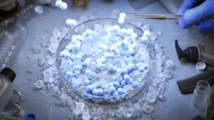
If successful, the implications of a quantum-enhanced electron microscope extend far beyond the laboratory. The most immediate impact would be in the field of structural biology. Understanding the exact 3D shape of proteins is essential for drug discovery; most modern medicines work by "docking" into a specific protein. Currently, scientists must freeze thousands of identical proteins in ice (Cryo-EM) and average their images to see them. A quantum microscope could potentially image individual proteins in a more natural state with far less preparation.
Furthermore, the technology could revolutionize material science. As the semiconductor industry moves toward 2-nanometer processes and beyond, the ability to inspect circuits without damaging them becomes critical. Quantum-enhanced imaging could allow for the inspection of delicate polymers and organic electronics that are currently difficult to study under electron beams.
"Quantum physics allows us to overcome the statistical limits that constrain conventional electron microscopes," says Pescoller. This statement marks a shift in how we view measurement itself. We are moving away from an era of "more power" and into an era of "smarter information."
Analysis of the Technological Shift
The development of the quantum electron microscope represents a maturation of the "Quantum 2.0" revolution. While the first quantum revolution gave us the transistor and the laser, the second revolution is focused on actively manipulating entanglement and superposition to perform tasks that were previously thought to be physically impossible.
By treating the electron beam as a data carrier rather than just a light source, the Austrian consortium is treating the microscope as a communication channel. In this framework, the goal is to maximize the "information per electron." This information-centric approach is likely to influence other fields of measurement, such as LIGO’s gravitational wave detection or MRI medical imaging, where quantum "squeezing" and entanglement are also being explored to reduce noise.
As the prototype at TU Wien nears completion, the scientific community awaits the first images. If the mathematical proofs translate into experimental reality, the "quantum eye" will provide a window into the building blocks of life that has, until now, remained shrouded in the noise of the subatomic world. The project stands as a testament to the power of interdisciplinary collaboration, proving that when quantum computing meets classical microscopy, the result is a vision of the future that is clearer than ever before.