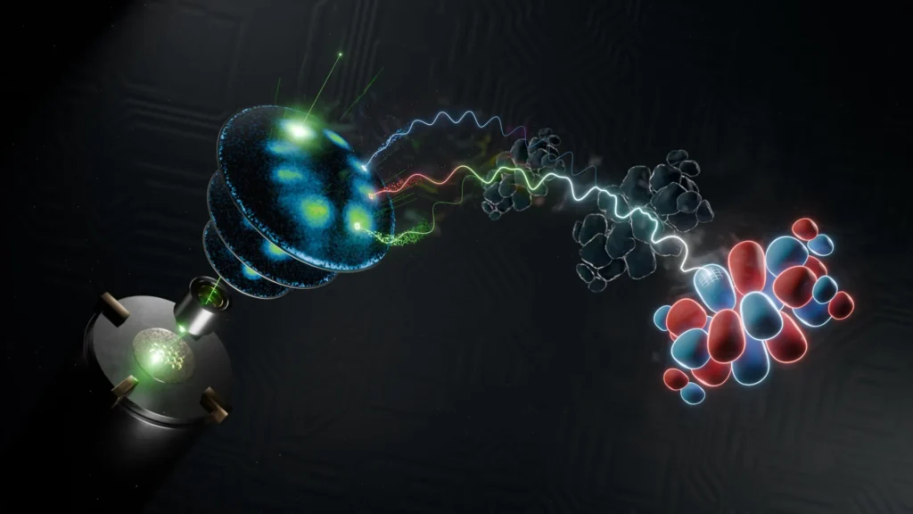
In a landmark achievement for the field of quantum chemistry, an interdisciplinary research team at the University of Göttingen has successfully produced a three-dimensional image of the wavefunction of an organic molecule. This breakthrough, recently detailed in the journal Nature Communications, represents a significant leap forward in our ability to visualize the fundamental building blocks of matter at the sub-atomic scale. By combining advanced photoelectron spectroscopy with sophisticated mathematical reconstruction algorithms, the researchers have managed to bypass long-standing limitations that previously required the use of massive, multi-billion-dollar synchrotron facilities. This development not only provides a clearer picture of molecular structures but also paves the way for "femtosecond videography," which would allow scientists to observe chemical reactions and electronic transitions in real-time at the speed of a quadrillionth of a second.
The Quantum Nature of Molecular Structures
To understand the magnitude of this achievement, one must first consider the counterintuitive nature of the quantum world. In classical physics, an object occupies a specific, measurable position in space. However, at the scale of electrons and atoms, this certainty vanishes. According to the principles of quantum mechanics, fundamental particles like electrons do not exist as discrete points but are instead described by a "wavefunction." This mathematical entity defines the probability distribution of a particle’s properties, such as its position, momentum, and spin.
Within the context of a molecule, these electron wavefunctions are referred to as "molecular orbitals." These orbitals are the "blueprints" of chemical behavior; they dictate how a molecule will bond with others, how it will react to light, and how it will conduct electricity. Until now, capturing a full three-dimensional representation of these orbitals has been an elusive goal for the scientific community. While two-dimensional projections have been possible, the transition to 3D imaging has historically been hampered by the "phase problem"—the inherent difficulty in measuring both the amplitude and the phase of a quantum wave simultaneously.
Methodology: Spectroscopy Meets Advanced Computation
The Göttingen team, led by Professor Stefan Mathias and Dr. Matthijs Jansen, approached this challenge by utilizing an indirect measurement technique known as photoelectron spectroscopy. In this process, a molecule is hit with high-energy light, causing it to eject electrons. By measuring the momentum of these emitted electrons, researchers can capture what is effectively "half" of the information contained within the wavefunction. Specifically, they can determine the magnitude of the momentum, but the spatial phase—the information regarding the wave’s relative timing and direction—is typically lost during the measurement process.
To recover this lost data, the team developed a revolutionary computational approach. Instead of relying solely on experimental measurements to fill in the gaps, they utilized a redesigned mathematical algorithm capable of reconstructing the missing phase information. This algorithm works by iteratively testing potential wavefunction structures against the measured data until a perfect match is found.
One of the most significant aspects of this new methodology is its efficiency. Traditionally, attempting to map a 3D orbital required an enormous amount of data, often necessitating weeks of measurement time at large-scale synchrotron radiation sources. The Göttingen team’s algorithm, however, is designed to be highly "data-light," requiring significantly fewer experimental data points to produce a reliable three-dimensional image. This efficiency is what allowed the team to move the experiment from a specialized national facility into a standard laboratory setting.
The Role of Laboratory-Based Soft X-Ray Sources
The second pillar of this breakthrough is the use of a high-performance, laboratory-based soft X-ray source. Historically, the high-energy photons required to probe molecular orbitals could only be produced by synchrotrons—massive particle accelerators that occupy entire building complexes. These facilities are expensive, oversubscribed, and often located far from the primary research laboratories.
By utilizing a compact, lab-based source that generates ultrashort soft X-ray pulses, the Göttingen researchers have "democratized" the process of 3D wavefunction imaging. This specific light source produces pulses of light that are not only high in energy but also incredibly brief. These short pulses are essential for capturing "snapshots" of electrons before the molecular structure has time to shift or vibrate. The combination of this localized X-ray technology and the new reconstruction algorithm creates a synergistic effect that makes 3D imaging both practical and accessible for a wider range of research institutions.
A Chronology of Quantum Imaging Progress
The journey toward 3D wavefunction imaging has been decades in the making, involving a series of incremental steps in both physics and mathematics:
- Early 20th Century: The development of the Schrödinger equation establishes the theoretical framework for wavefunctions and molecular orbitals.
- 1960s-1980s: The maturation of photoelectron spectroscopy allows scientists to begin measuring electron binding energies, though spatial mapping remains limited.
- 2000s: The emergence of "Photoemission Tomography" allows for the 2D reconstruction of molecular orbitals of flat molecules on surfaces, primarily using synchrotron radiation.
- 2010s: Researchers begin attempting to extend tomography to 3D, but are met with the "data bottleneck," where the required measurement time makes the process impractical for complex or changing systems.
- Present Day: The University of Göttingen team successfully integrates lab-based X-ray sources with phase-retrieval algorithms, achieving sub-atomic resolution in three dimensions for an organic molecule.
Technical Data and Resolution Benchmarks
The precision achieved in this study is noteworthy even by the standards of modern nanotechnology. The researchers were able to distinguish features within the molecular orbital that are smaller than the distance between the carbon atoms that form the molecule’s backbone. This level of resolution—often measured in picometers (one-trillionth of a meter)—allows scientists to see the subtle "lobes" and "nodes" of the electron cloud where chemical bonding occurs.
By imaging an organic molecule only a few nanometers in size, the team demonstrated that their technique is sensitive enough to handle the complex, non-symmetrical structures that are common in organic chemistry and biology. This is a significant departure from previous experiments that often focused on simpler, highly symmetrical inorganic crystals.
Official Responses and Scientific Impact
The implications of this research have been met with enthusiasm across the scientific community. Dr. Wiebke Bennecke, the study’s first author, emphasized the transformative potential of the time-resolution aspect of their work. "This technique might mean that stroboscopic videography becomes a reality," Bennecke stated. She noted that the ability to observe wavefunctions at femtosecond resolution would allow researchers to see how a molecule adapts to electronic or chemical changes in real-time.
Professor Stefan Mathias highlighted the fundamental shift in how quantum mechanics is studied. "The wavefunction is a fundamental quantity in quantum mechanics, yet it cannot be directly observed or measured," he explained. By creating a reliable proxy through reconstruction, the team has provided a new "lens" through which the quantum world can be viewed.
Inferred reactions from the broader chemistry and physics communities suggest that this technique will be particularly valuable for "orbital engineering." This field focuses on manipulating the shape and energy of molecular orbitals to create new materials with specific properties, such as high-efficiency solar cells, faster semiconductors, or more effective catalysts for industrial chemistry.
Analysis: Toward Ultrafast 3D Molecular Movies
The most profound implication of the Göttingen study lies in the future of "femtosecond videography." Currently, most images of molecular structures are static; they show the molecule in its ground state or a stable equilibrium. However, the most important processes in nature—such as photosynthesis, vision, and DNA repair—involve molecules that are in a state of constant, rapid flux.
A femtosecond is one-quadrillionth of a second. To put this in perspective, a femtosecond is to a second what a second is to about 32 million years. At this timescale, the motion of electrons can be captured as they jump from one atom to another during a chemical reaction. By applying the Göttingen team’s 3D imaging technique to these ultrafast timescales, scientists could essentially create "3D movies" of chemical bonds breaking and forming.
This capability would move the field of chemistry from a descriptive science to a truly predictive one. If researchers can see exactly how a wavefunction deforms when hit by a photon, they can design molecules that are better at harvesting light or resisting degradation. In the pharmaceutical industry, watching how a drug molecule’s wavefunction interacts with a protein’s wavefunction could lead to the development of medicines with fewer side effects and higher precision.
Broader Implications for Material Science and Quantum Computing
Beyond the realm of chemistry, this breakthrough has significant consequences for the development of next-generation technology. In the field of material science, understanding the 3D distribution of electrons is vital for creating "topological insulators" and other exotic states of matter that could revolutionize electronics.
Furthermore, as the world moves toward quantum computing, the ability to accurately map and verify the state of a quantum system is paramount. The Göttingen team’s reconstruction algorithm offers a potential pathway for verifying the "qubits" in a quantum processor, ensuring that they are in the correct state before a calculation begins.
The transition from synchrotrons to laboratory-based imaging also represents a shift toward more sustainable and rapid scientific discovery. When a researcher can test a hypothesis in their own lab rather than waiting months for a "beamtime" slot at a national facility, the pace of innovation accelerates exponentially.
Conclusion
The successful 3D imaging of a molecular wavefunction at the University of Göttingen marks the beginning of a new era in quantum observation. By solving the dual challenges of the "phase problem" and the "data bottleneck," the research team has turned a theoretical mathematical concept into a visible, actionable reality. As this technique is refined and applied to dynamic, time-resolved studies, the scientific community stands on the verge of witnessing the secret, high-speed life of molecules, potentially unlocking new ways to control the physical world at its most fundamental level. The move toward lab-based, high-resolution 3D imaging ensures that this powerful tool will soon be at the fingertips of researchers worldwide, driving advancements in everything from green energy to molecular medicine.