A groundbreaking study led by researchers at MIT has unveiled a novel method for precisely engineering the growth patterns of new blood vessels, a critical advancement with profound implications for regenerative medicine and tissue engineering. Published in the Proceedings of the National Academy of Sciences, the research demonstrates that the simple mechanical action of repeatedly jostling or stretching an artery can stimulate it to sprout new, smaller capillaries. Furthermore, the direction and degree of this mechanical stimulation can be meticulously controlled to direct the growth of these nascent vessels, offering unprecedented control over vascular network formation.
This discovery represents a significant leap forward in addressing one of the most persistent challenges in tissue engineering: the inability to reliably create organized, functional blood vessel networks within engineered tissues. Such networks are essential for the survival and integration of any implanted tissue or organ, ensuring the delivery of vital nutrients and oxygen while removing waste products.
The Critical Role of Vascularization in Tissue Engineering
For decades, the promise of regenerative medicine has been tempered by the immense difficulty of vascularizing engineered tissues. While scientists have made remarkable progress in growing various cell types and even rudimentary organs in laboratories, scaling these constructs to a clinically relevant size or complexity has often been thwarted by the lack of an integrated blood supply. Without a robust vascular network, cells beyond a few hundred micrometers from the tissue’s surface quickly suffer from hypoxia (lack of oxygen) and nutrient deprivation, leading to cell death and tissue failure. This limitation has been a primary bottleneck for developing functional artificial organs, large tissue grafts for reconstructive surgery, and effective models for drug testing.
Traditional approaches to encourage blood vessel growth, known as angiogenesis, have largely relied on chemical cues, such as growth factors like vascular endothelial growth factor (VEGF). While effective to some extent, these chemical methods often lack the precision needed to guide vessels into intricate, organized patterns. Distributing growth factors uniformly or in complex gradients within a three-dimensional tissue remains a formidable challenge, often resulting in chaotic and insufficient vascularization. Similarly, 3D printing technologies, while capable of fabricating larger vascular structures like arteries and veins, struggle to create the delicate, thread-like networks of capillaries—the microscopic vessels where nutrient and gas exchange truly occur. It is these fine capillary networks that are indispensable for tissue viability.
A Novel Mechanical Approach: "Moving is Good"
The research, spearheaded by Ritu Raman, an associate professor of mechanical engineering at MIT, and her team, introduces a paradigm shift by leveraging mechanical forces to orchestrate vascular growth. This approach draws inspiration from the body’s natural processes, where mechanical forces like blood flow and tissue deformation are known to play crucial roles in vascular development and remodeling. Raman’s laboratory has a history of exploring the impact of mechanical "exercise" on biological systems, having previously demonstrated its efficacy in guiding the growth of artificial muscles and nerves.
In their earlier work, the team developed a sophisticated "organ-on-a-chip" platform. This system involved embedding a small magnet within a nutrient-rich gel and then culturing living muscle or neuron cells on the gel’s surface. By manipulating an external magnet, they could repeatedly pull the embedded magnet and the cell-covered gel back and forth, effectively subjecting the cells to controlled mechanical strain. These experiments revealed that this physical "workout" directly influenced how the cells grew and organized, leading to more functional tissues.
Applying this established methodology to vascular engineering, the researchers constructed a "blood-vessel-on-a-chip." This miniature device, smaller than a postage stamp, contained a gel infused with nutrients and a tiny magnet. A thin, hollow channel was created lengthwise through the gel and coated with live endothelial cells. Endothelial cells are the specialized cells that line the inside of blood vessels and are responsible for forming new capillaries through a process called sprouting angiogenesis. Once the cells adhered to the channel’s surface and formed a nascent "main artery," they began to spontaneously sprout new, capillary-like vessels into the surrounding gel.
To investigate the effect of mechanical stimulation, the device was placed under a motorized stage equipped with suspended magnets. The researchers then systematically moved these magnets back and forth, varying both the direction and the degree of displacement. The ensuing observations were striking and formed the core of their breakthrough.
Precise Control Over Vascular Architecture
The experiments revealed a clear and direct correlation between mechanical stimulation and blood vessel growth. When the central artery was left undisturbed in the gel, it did produce some new vessels, but their growth was largely random and unorganized. However, when the artery was subjected to repeated jostling, a significantly greater number of capillaries sprouted from its length.
The team meticulously quantified these effects:
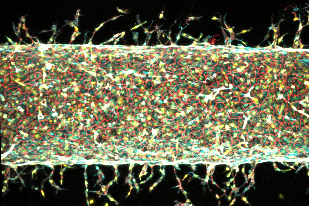
- Enhanced Sprouting: A gentle stretch, equivalent to 5 percent of the gel’s total width, dramatically increased the number of new vessels emerging from the main artery.
- Influenced Length: A more significant stretch, around 15 percent of the gel’s width, resulted in fewer new vessels overall, but those that did sprout grew considerably longer. This suggests a potential trade-off between vessel density and length, which could be precisely tuned for different applications.
- Directed Growth: Perhaps most critically, by changing the direction of the mechanical stretching, the researchers could effectively "steer" the growth of the new vessels. The capillaries consistently followed the pattern of the applied mechanical stimulation, making turns and extending in alignment with the induced strain.
As Professor Raman succinctly put it, "Moving is good, which is always the takeaway of everything we do in our lab. Mechanical forces play an important role in our bodies. That means that if you want to grow more or less vessels, or shorter or longer vessels, or vessels in certain directions, we now know how to do that." This statement underscores the unprecedented level of control this method offers over the traditionally unruly process of angiogenesis.
Unveiling the Genetic Mechanism: The Role of PIEZO1
Beyond demonstrating the efficacy of mechanical stimulation, the researchers delved into the underlying biological mechanisms. This deeper understanding is crucial for validating the findings and potentially optimizing the protocol. Their investigation led them to the PIEZO1 gene.
The inspiration for this line of inquiry came from Ardem Patapoutian, a molecular biologist who was awarded the Nobel Prize in Physiology or Medicine in 2021. Patapoutian’s seminal work identified ion channels in cell membranes—specifically PIEZO1 and PIEZO2—that act as mechanosensors. These channels open and close in response to mechanical pressure, controlling the flow of ions into and out of cells, thereby translating physical forces into biochemical signals. Essentially, they are the cell’s gatekeepers for mechanical stimuli.
Raman had the opportunity to present her team’s preliminary findings on blood vessel growth under mechanical stimulation to Patapoutian. He hypothesized that the observed angiogenesis could be mediated by the PIEZO1 channel. The repeated mechanical exercise of the central artery might be activating these channels in the endothelial cells, thereby triggering the downstream signaling pathways that lead to new blood vessel formation.
To test this hypothesis, Raman’s team employed gene-editing techniques to suppress the activity of the PIEZO1 gene in the endothelial cells. The results were compelling: when the PIEZO1 gene was less active, significantly fewer new blood vessels sprouted from the central artery, even when it was subjected to the same mechanical exercise. This direct evidence confirmed that PIEZO1 acts as a crucial molecular transducer, linking mechanical forces to the biological response of angiogenesis. This discovery not only provides a mechanistic explanation for their findings but also opens avenues for pharmacological interventions that could modulate PIEZO1 activity to either enhance or inhibit vascular growth as needed.
Transformative Potential in Regenerative Medicine and Beyond
The implications of this research are far-reaching and could revolutionize several fields:
- Engineered Organs and Tissues: The ability to program organized blood vessel networks is a monumental step towards creating functional, implantable artificial organs. Imagine a future where a failing heart, kidney, or liver could be partially or fully replaced with bioengineered counterparts, complete with their own integrated vascular supply. This research brings that vision closer to reality. For instance, in cases of myocardial infarction (heart attack), where heart tissue is damaged due to lack of blood flow, this technology could facilitate the growth of new, healthy, vascularized tissue to restore cardiac function.
- Tissue Repair and Regeneration: For patients suffering from severe injuries, burns, or debilitating diseases like diabetes that impair wound healing and blood flow (e.g., diabetic foot ulcers, peripheral artery disease), this method could be used to regenerate healthy, vascularized tissue. It could also improve the success rate of large tissue grafts by ensuring their immediate and robust integration into the host’s circulatory system.
- Disease Modeling and Drug Discovery: The "blood-vessel-on-a-chip" platform, with its unprecedented control over vascular architecture, offers a superior in vitro model for studying various vascular diseases, such as atherosclerosis, vasculitis, and tumor angiogenesis. Researchers could precisely mimic diseased vascular environments and test the efficacy of new drugs or therapies with greater accuracy than current models allow. This could significantly accelerate drug discovery for conditions like cancer, where inhibiting tumor angiogenesis is a key therapeutic strategy.
- Understanding Biological Processes: Beyond direct therapeutic applications, this research deepens our fundamental understanding of how mechanical forces influence cellular behavior and tissue development. This knowledge could have implications for fields ranging from developmental biology to exercise physiology.
Co-author Jessica Shah highlighted the immediate next steps: "We are now investigating how precisely patterning blood vessel growth can help improve muscle function." This suggests a direct progression towards integrating these engineered vascular networks into larger functional tissues, such as muscle constructs, to evaluate their efficacy in restoring biological function.
Broader Impact and Future Outlook: Challenges and Opportunities
While the findings are incredibly promising, it is important to acknowledge that this is foundational research. Several challenges lie ahead before these discoveries can translate into clinical applications.
- Scaling Up: The current "blood-vessel-on-a-chip" is a small-scale laboratory model. Scaling up the production of complex, vascularized tissues and organs for human implantation will require significant engineering innovation. This includes developing bioreactors that can maintain appropriate mechanical stimulation and nutrient flow for larger constructs.
- Integration with Host Tissue: Once implanted, engineered vascular networks must seamlessly integrate with the host’s existing circulatory system. This involves complex biological signaling and physical connections that need to be understood and controlled. Immunological compatibility will also be a key consideration for long-term success.
- Long-Term Viability and Functionality: Ensuring that the engineered vessels remain stable, functional, and capable of adapting to physiological demands over extended periods is crucial.
- Regulatory Pathways: The path to clinical approval for novel bioengineered tissues is rigorous and lengthy, requiring extensive preclinical testing and human clinical trials to demonstrate safety and efficacy.
Despite these challenges, the scientific community is likely to react to this research with considerable excitement. Experts in regenerative medicine have long sought such precise control over vascularization. This work provides a powerful new tool that complements and potentially surpasses existing methods. It shifts the paradigm from purely chemical or passive structural cues to active, dynamic mechanical regulation, mimicking the sophisticated interplay of forces that govern biological development in vivo.
The research was supported, in part, by critical funding from the U.S. Department of War Army Research Office Early Career Program and PECASE Grant, and a Department of War DURIP Program Grant. This underscores the strategic importance of such advancements for military medicine, particularly in treating traumatic injuries where tissue regeneration and vascular repair are paramount.
In conclusion, the MIT team’s discovery that mechanical stimulation can precisely program blood vessel growth marks a pivotal moment in tissue engineering. By harnessing the power of physical forces and elucidating the underlying genetic mechanisms involving PIEZO1, they have opened a new frontier in the quest to build functional biological tissues and organs. While the journey to clinical translation will be long, this breakthrough offers a robust and elegant solution to one of regenerative medicine’s most enduring puzzles, bringing humanity closer to a future where debilitating diseases and injuries can be effectively treated with engineered biological replacements.