In a significant leap forward for cellular biology and biomedical research, scientists at the Broad Institute of MIT and Harvard, in collaboration with MIT, have developed an innovative "cellular self-reporting" approach that enables the continuous monitoring of a living cell’s transcriptome without causing cellular damage or death. This breakthrough method, detailed in the prestigious journal Cell, promises to fundamentally alter how researchers study cellular identity, genetic activity, disease progression, and drug responses by providing dynamic, real-time insights into gene expression previously unattainable.
The Limitations of Traditional Transcriptomics: A Snapshot in Time
For years, the scientific community has relied on various methods to measure a cell’s transcriptome—the complete set of RNA molecules produced by a cell at a given time. This transcriptome serves as a vital blueprint, reflecting which genes are active and to what extent, thereby revealing a cell’s identity, function, and response to its environment. Understanding the transcriptome is crucial for unraveling the complexities of biological processes, from development and differentiation to disease pathogenesis and therapeutic intervention.
However, the prevailing methodologies for transcriptomic analysis have inherent limitations. Almost universally, these techniques necessitate the lysis, or destruction, of the cell to extract its RNA content. This fundamental requirement means that researchers can only obtain a single "snapshot" of a cell’s genetic activity at a specific moment. While valuable, these static observations fail to capture the dynamic changes in gene expression that occur as cells mature, respond to stimuli, or transition through various states, such as those associated with disease progression or drug treatment. Imagine trying to understand a complex movie by only viewing isolated frames; the plot, character development, and intricate interactions would be largely lost. This is precisely the challenge posed by traditional transcriptomics when studying biological systems that are inherently dynamic and evolving. The inability to track the same cell or cell population over time has been a significant bottleneck, particularly in longitudinal studies of disease models, developmental biology, and drug pharmacodynamics.
The Breakthrough: Unlocking Continuous Live-Cell Monitoring
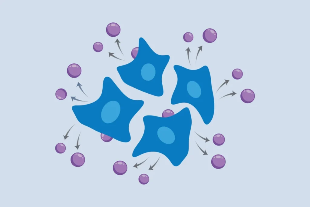
The new "cellular self-reporting" method elegantly bypasses these limitations by allowing living cells to autonomously share their transcriptomic information. Instead of researchers actively extracting RNA through destructive means, the engineered cells themselves package and deliver their RNA into the surrounding culture medium. Scientists can then simply sample this medium repeatedly, isolating the secreted RNA to analyze the transcriptome without ever disturbing or harming the cells. This non-invasive, longitudinal capability marks a paradigm shift, enabling researchers to observe how gene activity within the same cell population evolves over extended periods, offering unprecedented insights into cellular biology.
Paul Blainey, a core member of the Broad Institute and a professor of biological engineering at MIT, and the senior author of the study, emphasized the transformative potential of this work. "Our lab focuses our time and resources on developing tools that will actually get used and make real impact on the broader field," Blainey stated. "It’s so gratifying to see a real coming to fruition of this concept, which was complete science fiction when we started. It’s a great example of the innovative impact long-term high-risk, high-reward research can have." His remarks underscore the years of dedicated effort and visionary thinking required to translate such an ambitious concept into a tangible, functional technology.
A Decade in the Making: The Genesis of a Visionary Idea
The journey to develop this revolutionary method spans more than a decade, rooted in a persistent challenge that Paul Blainey’s lab aimed to resolve: how to perform RNA sequencing without sacrificing the cells being studied. "The existing methods were a bit medieval and involved stabbing cells or cutting pieces off of them," Blainey recounted, highlighting the often-brutal nature of previous approaches. This dissatisfaction with the status quo fueled a quest for a more refined, less invasive technique.
Inspired by the remarkable success and widespread adoption of molecular technologies such as CRISPR-based gene editing, Blainey and study first author Jacob Borrajo committed to developing a purely molecular method. They recognized that while this path would be fraught with technical complexities and demand significant time and resources, a molecularly encoded solution would ultimately be more scalable, robust, and accessible for the broader scientific community. This foresight was critical, as the ease of adoption is a key determinant of a new technology’s impact. Their vision was not just to solve a problem, but to create a tool that could be widely integrated into diverse research settings, democratizing access to dynamic transcriptomic data.
Mechanism of Innovation: Harnessing Viral Biology for Scientific Gain
The elegant solution developed by the Broad and MIT team draws inspiration from an unexpected source: retroviruses. These ubiquitous biological entities have, over millions of years of evolution, perfected the art of packaging their RNA genomes into protective protein shells, enabling their efficient spread from one infected cell to another. This natural viral mechanism provided the crucial conceptual framework for the "cellular self-reporting" system.
To implement their method, the researchers ingeniously engineered mammalian cells to express a specific retroviral structural protein. Crucially, this protein was designed not only to encapsulate viral RNA but also to promiscuously recruit and encapsulate the cell’s own messenger RNA (mRNA) and other RNA species. Once integrated into the cell’s membrane, this viral protein facilitates the formation of a protein shell around the captured cellular RNA, creating what are termed "virus-like particles" (VLPs). These VLPs, containing a representative sample of the cell’s transcriptome, then bud off from the cell membrane and are released into the surrounding liquid culture medium.
The subsequent steps are remarkably simple: scientists merely collect a sample of the culture medium, isolate the RNA contained within these VLPs, and then perform standard RNA sequencing. This process yields a comprehensive view of the cell population’s transcriptome at that specific time point, all accomplished without causing any damage or perturbation to the living cells. This non-destructive nature is paramount, as it allows for repeated sampling from the same cell population, providing an unprecedented temporal resolution of gene expression dynamics.
Mohamad Najia, a co-first author and research fellow in the Blainey lab and George Daley’s lab at Boston Children’s Hospital, underscored the practical advantages of their approach. "Compared to methods using robotics or mechanical biopsies of cells, our molecularly encoded solution could be much more broadly enabling for the average life science or biomedical lab, particularly the time dynamic questions that we hope to elucidate with this technology," Najia stated. This sentiment was echoed by co-first author Anna Le, a postdoc in the Blainey lab, who also played a pivotal role alongside Borrajo and Najia in leading this groundbreaking work. Their emphasis on accessibility and scalability suggests a technology poised for widespread adoption, potentially democratizing complex longitudinal transcriptomic studies.
Validation Across Diverse Cellular Systems: Demonstrating Broad Applicability
A critical measure of any new scientific method is its versatility and robustness across various biological contexts. The Broad and MIT team rigorously tested their "cellular self-reporting" method, demonstrating its broad applicability across an impressive array of cellular models. They successfully applied the technique to:
- Immortalized human cell lines: Standard laboratory workhorses, validating the method’s fundamental functionality.
- Cancer cell lines: Crucial for oncology research, enabling dynamic studies of tumor progression and drug resistance.
- Stem cells and their differentiated neuronal cells: Essential for developmental biology, regenerative medicine, and neurobiology, allowing insights into differentiation pathways and neuronal maturation.
- Primary cells from human donors: Representing more physiologically relevant models, confirming the method’s utility in systems closer to in vivo conditions.
Beyond single-cell type cultures, the researchers also tackled more complex scenarios. They successfully applied the method to a co-culture system comprising two distinct human cell types growing together. To differentiate between the transcriptomic signals from each cell type, they ingeniously incorporated unique molecular tags into the virus-like particles, allowing for the precise attribution of RNA to its cell of origin during analysis. This capability is vital for studying complex cellular interactions in heterogeneous tissues and organ models.
Furthermore, the "cellular self-reporting" system proved valuable for investigating systems where maintaining crucial three-dimensional structures is paramount. The team demonstrated its efficacy on spheroids of human endothelial cells—3D aggregates that mimic tissue architecture. They successfully captured short-term transcriptional changes following biochemical stimulation, showcasing the method’s ability to monitor responses in physiologically relevant 3D contexts without disrupting their intricate organization. This is a significant advantage over methods that require dissociation of such structures for single-cell analysis, which can introduce artifacts and lose spatial information.
Enhancing Complex Models: Organoids and Organ-on-a-Chip Technology
One of the most exciting applications of the new method lies in its potential to revolutionize research using advanced in vitro models like organoids and organ-on-a-chip devices. These sophisticated systems are engineered to mimic the physiology and pathology of human organs more accurately than traditional 2D cell cultures. They hold immense promise for minimizing preclinical animal testing, accelerating drug discovery, and advancing personalized medicine. However, their inherent complexity often makes retrieving cells for analysis challenging and disruptive, limiting the scope of longitudinal studies.
In a key collaboration, the Broad and MIT team partnered with Linda Griffith, a professor of biological and mechanical engineering at MIT, a pioneer in tissue engineering and organ-on-a-chip technology. Applying the "cellular self-reporting" method to Griffith’s lab’s organ-on-a-chip devices, the researchers were able to monitor gene expression dynamics in endothelial cells within these complex microfluidic systems over time. This capability revealed fascinating insights, such as changes in genes related to how tissues form vascular networks. Crucially, these transcriptional patterns were found to depend on the source of supporting fibroblasts—whether derived from the uterus or the lung—highlighting the method’s power to uncover subtle yet significant biological differences in a dynamic, non-invasive manner. This represents a major step towards making organ-on-a-chip models even more powerful predictive tools, enabling the continuous assessment of drug efficacy, toxicity, and disease modeling in a human-relevant context.
Broader Scientific Implications and Transformative Potential
The introduction of "cellular self-reporting" transcriptomics carries profound implications across a multitude of scientific and medical disciplines:
- Disease Mechanisms: Researchers can now track the precise genetic changes that occur as cells transition from healthy to diseased states, providing unprecedented insights into the initiation and progression of conditions like cancer, neurodegenerative disorders (e.g., Alzheimer’s, Parkinson’s), and metabolic diseases. This temporal resolution will be invaluable for identifying early biomarkers and critical intervention points.
- Drug Discovery and Development: The ability to continuously monitor how cells respond to drug candidates over time will accelerate preclinical drug screening. Scientists can assess drug efficacy, identify potential off-target effects, monitor the development of drug resistance, and optimize dosing regimens in real-time, leading to more effective and safer therapeutics. This could significantly reduce the time and cost associated with bringing new drugs to market.
- Developmental Biology: Understanding cell differentiation, tissue formation, and organ development requires observing dynamic gene expression patterns. This method offers a powerful tool to track these processes in living systems, shedding light on the intricate genetic programs that orchestrate life.
- Regenerative Medicine: For stem cell therapies, monitoring the differentiation and maturation of transplanted cells in vitro or in animal models without disturbing them is critical for ensuring their safety and efficacy.
- Toxicology: Assessing the cellular response to environmental toxins or chemical exposures over time can provide a more comprehensive understanding of their impact, moving beyond single-point measurements.
- Personalized Medicine: In the future, it might be conceivable to use patient-derived cells in in vitro models to test various treatments and monitor their individual responses in real-time, tailoring therapies more precisely to each patient’s genetic makeup and disease profile.
The non-invasive nature of this technology also aligns with the growing emphasis on 3R principles (Replace, Reduce, Refine) in animal research, potentially allowing for more sophisticated in vitro models that reduce the reliance on animal testing while providing richer data.
Future Directions and Unanswered Questions
The Broad team is far from resting on its laurels. They are actively exploring new applications and biological questions that can be addressed with their innovative system. A key area of ongoing development is to enhance the approach’s feasibility for studying single cells. While the current method provides population-level transcriptomic data, achieving single-cell resolution in a continuous, non-invasive manner would represent another monumental leap, allowing researchers to track the unique trajectories of individual cells within a heterogeneous population. This would be particularly impactful for understanding cellular plasticity, rare cell types, and the stochastic nature of gene expression.
For now, the researchers are optimistic that their "cellular self-reporting" method will be readily adopted by scientists interested in gaining a deeper, more dynamic understanding of how cells and tissues change over time. The potential for this technology to unravel previously hidden biological processes and accelerate the development of new therapies is immense, marking a pivotal moment in the ongoing quest to decipher the intricate language of life encoded in our genes. The "message in a bottle" delivered by these self-reporting cells promises to unlock secrets that have long remained beyond the reach of conventional scientific inquiry.