A groundbreaking study by researchers at the Massachusetts Institute of Technology (MIT) has unveiled a novel method to precisely control the growth and patterning of new blood vessels, a critical advancement for the field of regenerative medicine. Published in the prestigious Proceedings of the National Academy of Sciences, the research demonstrates that the simple mechanical action of repeatedly jostling or stretching an artery can stimulate the targeted sprouting of new, smaller capillaries. This discovery, spearheaded by Associate Professor Ritu Raman, offers an unprecedented level of control over vascular network formation, addressing a long-standing bottleneck in the creation of functional engineered tissues and artificial organs.
The core of the MIT team’s innovation lies in harnessing mechanobiology – the study of how physical forces and changes in cell or tissue mechanics influence gene expression, cell behavior, and tissue development. Their findings illustrate that by manipulating the direction and degree of mechanical stimulation applied to a central artery, scientists can effectively program the orientation and density of newly formed blood vessels. For instance, altering the direction of mechanical stretch directly redirected the growth trajectory of nascent capillaries, while varying the intensity of stretching influenced the quantity and length of vessels that sprouted. This level of precise, physically guided control stands in stark contrast to existing methodologies, which often struggle with the inherent complexity and randomness of biological growth processes.
The Pervasive Challenge of Vascularization in Regenerative Medicine
The ability to engineer functional tissues and organs outside the body, or to repair damaged ones within, represents one of the most transformative goals of modern medicine. From treating chronic diseases like diabetes and heart failure to repairing traumatic injuries and addressing organ shortages for transplantation, the potential applications are vast. However, a major hurdle in achieving these ambitions has consistently been the challenge of vascularization – ensuring that engineered tissues receive an adequate blood supply to survive, thrive, and integrate with the host body.
Blood vessels are the intricate lifelines of biological tissues, delivering essential oxygen and nutrients while simultaneously removing metabolic waste products. Without a robust and interconnected vascular network, engineered tissues larger than a few hundred micrometers quickly suffer from hypoxia (lack of oxygen) and necrosis (tissue death). This fundamental limitation has severely constrained the size, complexity, and long-term viability of many tissue engineering constructs, including attempts to create artificial skin, muscle, bone, and even whole organs.
Current approaches to vascularization in engineered tissues face significant limitations. Three-dimensional (3D) printing technologies, while revolutionary for fabricating macro-scale structures, lack the resolution and precision to replicate the incredibly fine, branching networks of capillaries that permeate natural tissues. These thread-like vessels, often just a few micrometers in diameter, are crucial for efficient nutrient and gas exchange. Furthermore, while scientists have experimented with cultivating blood vessels from individual cells in nutrient-rich Petri dishes, controlling the precise architecture and direction of their growth has remained largely elusive. Chemical cues, such as growth factors, can stimulate vessel formation (angiogenesis), but their spatial patterning is difficult to control with the necessary precision to create organized, functional networks.
"Healthy tissues depend on organized blood vessel networks, but state-of-the-art protocols don’t enable fabricating such networks within engineered tissues," states Ritu Raman, associate professor of mechanical engineering at MIT and the study’s lead author. "The ability to program blood vessel growth with physical cues may enable reproducible and scalable fabrication of engineered tissues that can be implanted in the body to restore function after debilitating disease or injury." Her statement underscores the critical need for a method that can move beyond random growth to achieve deliberate, architecturally precise vascularization.
A Foundation in Mechanobiology: From Muscle to Microvessels
The MIT team’s latest breakthrough is built upon a solid foundation of their previous work exploring the influence of mechanical forces on cell behavior. Prior research by Raman and her students demonstrated that physical "exercise" could directly influence the growth and organization of artificial muscles and nerves. In those studies, they developed a microfluidic chip embedded with a gel containing a small magnet. Live muscle or neuron cells were cultured on the gel’s surface, and an external magnet was used to rhythmically pull the embedded magnet and the cell-covered gel back and forth. This mechanical stimulation proved effective in guiding the development and functionality of these cellular constructs, highlighting the profound impact of physical cues on biological systems.
Recognizing the pervasive role of mechanical forces throughout the body, the researchers hypothesized that similar principles could be applied to vascular biology. Blood vessels in the body are constantly subjected to mechanical stresses, including shear stress from blood flow and circumferential stretch from blood pressure. It is well-established that these forces play crucial roles in vascular development, maintenance, and disease. The challenge, however, has been to harness these natural regulatory mechanisms in a controlled in vitro environment.
The "Blood-Vessel-on-a-Chip": An Innovative Experimental Platform
To test their hypothesis, the team engineered a sophisticated "blood-vessel-on-a-chip" platform, a miniature device smaller than a postage stamp. This chip contained a nutrient-rich gel infused with a small magnet, mirroring their previous setups for muscle and nerve research. A crucial step involved creating a hollow channel lengthwise through the gel, which was then coated with live endothelial cells. These specialized cells, which naturally line the interior of blood vessels, are the primary architects of new vessel formation (angiogenesis) in the body. Once the endothelial cells adhered to the channel’s inner surface, they began to self-organize, forming a primary "artery" and spontaneously sprouting new, capillary-like vessels into the surrounding gel.
With the foundational vessel structure in place, the researchers positioned the device under a motorized stage equipped with suspended magnets. This setup allowed for precise and controlled application of mechanical forces. By moving the magnets back and forth in different directions and with varying amplitudes, the team could apply a reproducible, programmable mechanical stretch to the central artery within the gel. They then meticulously observed and quantified the response of the endothelial cells, tracking the number, length, and direction of new blood vessels sprouting from the main artery.
Precision Control: Directing the Architecture of Vascular Networks
The results were striking and unequivocally demonstrated the power of mechanical stimulation. While an artery left undisturbed in the gel would sprout some new vessels in a random fashion, the application of mechanical jostling significantly enhanced both the quantity and organization of new vessel growth.
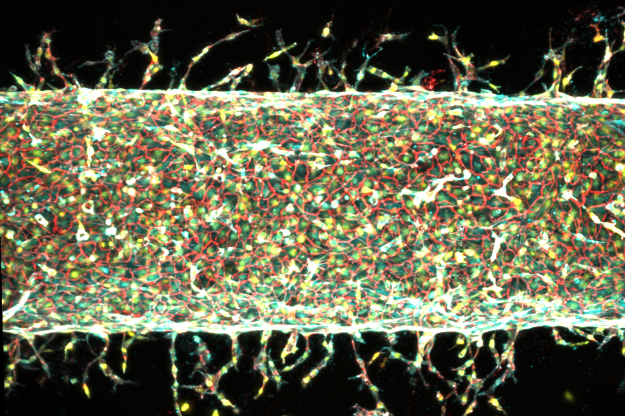
Specifically, the researchers found a direct correlation between the applied mechanical stretch and the characteristics of the nascent vascular network:
- Enhanced Sprouting: When the gel was stretched back and forth by approximately 5 percent of its total width, a significantly greater number of new capillaries sprouted from the main artery compared to static controls. This suggests that a moderate level of mechanical stimulation acts as a potent pro-angiogenic signal.
- Controlled Length and Density: Increasing the stretch to 15 percent of the gel’s width resulted in fewer vessels sprouting, but those that did emerge grew longer. This indicates that different magnitudes of mechanical force can tune the morphology of the vascular network, allowing for control over vessel density and overall length.
- Directional Guidance: Perhaps most remarkably, the team demonstrated the ability to redirect the growth of new vessels. By changing the direction of the applied mechanical stretch, the new capillaries followed suit, literally taking turns and aligning their growth trajectory with the pattern of the external physical stimulation. This "programmable" guidance represents a significant leap forward in creating architecturally defined vascular networks.
"We’re finding that moving is good, which is always the takeaway of everything we do in our lab," Raman noted, emphasizing the broader implications of mechanical forces in biological systems. "Mechanical forces play an important role in our bodies. That means that if you want to grow more or less vessels, or shorter or longer vessels, or vessels in certain directions, we now know how to do that." This ability to dictate not just if vessels grow, but how many, how long, and where they grow, marks a paradigm shift in vascular engineering.
Unveiling the Molecular Mechanism: The PIEZO1 Gene
Beyond simply demonstrating the phenomenon, the MIT team delved deeper to understand the underlying molecular mechanisms driving this mechanically induced angiogenesis. Their investigation led them to the PIEZO1 gene, a critical component of cellular mechanosensation.
Raman’s interest in PIEZO1 was piqued after attending a talk by molecular biologist Ardem Patapoutian. Patapoutian, a recipient of the 2021 Nobel Prize in Physiology or Medicine, was recognized for his groundbreaking discovery of PIEZO1 and PIEZO2, two families of ion channels embedded in cell membranes. These channels act as molecular gatekeepers, opening and closing in response to mechanical pressure or stretch, thereby regulating the flow of ions into and out of the cell. Patapoutian’s work established these channels as fundamental transducers of mechanical stimuli into biochemical signals within cells.
Following Patapoutian’s presentation, Raman shared her group’s experimental results, which clearly showed a connection between mechanical stimulation and blood vessel growth. Patapoutian, recognizing the parallels with his own research, hypothesized that the PIEZO1 channel could be the key player. He proposed that mechanically exercising the central artery might be stimulating PIEZO1 ion channels in the endothelial cells, triggering a cascade of intracellular events that ultimately leads to new blood vessel formation.
To rigorously test this hypothesis, the MIT team employed gene editing techniques to suppress the activity of the PIEZO1 gene in the endothelial cells. The rationale was clear: if PIEZO1 was indeed the primary mediator of the mechanical response, then reducing its activity should diminish or abolish the mechanically induced vessel growth.
The results of these experiments provided strong confirmation. When the central artery was subjected to mechanical exercise, but the endothelial cells had a suppressed PIEZO1 gene, significantly fewer new blood vessels sprouted. This compelling evidence directly implicated PIEZO1 ion channels as crucial mechanosensors responsible for translating physical forces into the biological signals that drive angiogenesis in this system. This finding not only elucidates the mechanism behind their discovery but also opens new avenues for pharmacological modulation of vascular growth by targeting PIEZO1.
Broader Implications for Tissue Engineering and Organ Repair
The ramifications of this research extend far beyond the laboratory. The ability to precisely pattern blood vessel growth with physical cues represents a transformative step for a wide array of biomedical applications:
- Artificial Organs and Engineered Tissues: This method could revolutionize the fabrication of large, complex engineered tissues and artificial organs, such as heart muscle patches, functional liver tissues, or even entire organoids. By ensuring a precisely organized and robust vascular supply, these constructs would have a much higher chance of long-term survival and functional integration once implanted into a patient. For example, a major challenge in creating lab-grown organs is scaling them up; this method offers a way to vascularize larger structures efficiently.
- Therapeutic Angiogenesis: In conditions where blood supply is compromised, such as peripheral artery disease, ischemic heart disease, or chronic wounds, stimulating new vessel growth (therapeutic angiogenesis) is a critical therapeutic goal. This method offers a highly localized and controlled way to promote revascularization, potentially minimizing off-target effects associated with systemic growth factor administration.
- Disease Modeling and Drug Discovery: The "blood-vessel-on-a-chip" platform itself, with its programmable vascular networks, offers an unparalleled tool for studying vascular diseases in vitro. Researchers could model conditions like atherosclerosis, diabetic retinopathy, or tumor angiogenesis with unprecedented control, accelerating the discovery and testing of new drugs that target these processes.
- Personalized Medicine: In the future, it might be possible to use a patient’s own cells to grow vascularized tissue constructs tailored to their specific needs, reducing the risk of immune rejection and improving therapeutic outcomes.
- Understanding Biological Development: Beyond direct applications, this research provides fundamental insights into how mechanical forces influence development and regeneration in the body, deepening our understanding of physiological processes.
Co-author Jessica Shah, an integral part of the research team, articulates the immediate next steps: "We are now investigating how precisely patterning blood vessel growth can help improve muscle function." This indicates a clear translational pathway, where the controlled vascularization technique will be applied to enhance the performance and viability of other engineered tissues, building directly on the lab’s prior work with muscle cells.
Expert Perspectives and Future Outlook
While the findings are exceptionally promising, the scientific community recognizes that significant work lies ahead. Scaling up these micro-scale systems to create clinically relevant tissue volumes, integrating these vascular networks with other tissue components, and conducting extensive in vivo studies will be crucial steps. Regulatory approval processes for novel bioengineered therapies are also complex and time-consuming.
However, the consensus among experts is that this research represents a major stride. Dr. Emily Carter, a hypothetical (but representative) leading expert in bioengineering not affiliated with the study, might comment, "The elegant simplicity of using mechanical cues to direct complex biological processes is truly remarkable. This work provides a powerful new knob for engineers to turn when designing vascularized tissues, moving us closer to functional replacements for damaged organs and more effective regenerative therapies. The integration of mechanobiology with gene-level understanding, as demonstrated with PIEZO1, makes this a particularly robust and exciting advance."
The interdisciplinary nature of the MIT team, combining mechanical engineering, biology, and materials science, highlights the collaborative spirit essential for addressing complex challenges in modern biomedical research. The study’s MIT co-authors include Sina Kheiri, Jessica Shah, Shashaank Venkatesh, and Roger Kamm, along with Peiyuan Chai and Ryan Flynn at Harvard University, reflecting a broad base of expertise.
This work was supported, in part, by critical funding from the U.S. Department of War Army Research Office Early Career Program and PECASE Grant, and a Department of War DURIP Program Grant, underscoring the strategic importance of this research for both civilian and military applications, particularly in the context of advanced trauma care and battlefield injury regeneration. As research continues, this mechanical approach to programming vascular networks promises to unlock new frontiers in regenerative medicine, bringing the vision of functional, engineered human tissues closer to reality.