A groundbreaking study led by researchers at the Massachusetts Institute of Technology (MIT) has unveiled a novel method for cultivating and precisely controlling the growth patterns of new blood vessels, a critical advancement with profound implications for regenerative medicine, artificial organ development, and disease modeling. The team discovered that the simple mechanical action of repeatedly jostling or stretching an artery directly stimulates it to sprout smaller, intricate capillaries. Crucially, by manipulating the direction and degree of this mechanical stimulation, scientists can program the orientation and quantity of these new vessels. This pioneering work, detailed in the Proceedings of the National Academy of Sciences (PNAS), offers a transformative approach to engineering complex vascular networks that are essential for the viability and function of any living tissue or organ.
The Urgent Need for Engineered Vascular Networks
The ability to engineer functional tissues and organs has long been a holy grail in biomedical science. From repairing damaged heart tissue after a myocardial infarction to creating pancreatic islets for diabetes patients or entirely new organs for transplant, the promise of regenerative medicine is immense. However, a persistent and formidable challenge has been the lack of a robust, reliable method for creating intricate, hierarchical blood vessel networks that can adequately supply these engineered tissues with oxygen and nutrients, and remove waste products. Without an integrated vascular system, engineered tissues often fail to thrive, suffering from necrosis within days due as cells starve and suffocate.
Current approaches to vascularization have met with limited success. While advanced techniques like 3D bioprinting can produce larger arteries and veins, they struggle to replicate the delicate, micron-scale complexity of capillary networks that permeate every cell in the body. Another strategy involves culturing endothelial cells – the cells that line blood vessels – in nutrient-rich dishes with various growth factors. While this can induce vessel formation, controlling the precise architecture, density, and direction of growth has remained largely elusive. Chemical cues, while powerful, are difficult to pattern with the precision required to mimic the body’s natural vasculature.
"Healthy tissues depend on organized blood vessel networks, but state-of-the-art protocols don’t enable fabricating such networks within engineered tissues," explains Ritu Raman, an associate professor of mechanical engineering at MIT and co-lead author of the study. "We thus need other types of patternable cues that can help us build tissues with organized vessels. The ability to program blood vessel growth with physical cues may enable reproducible and scalable fabrication of engineered tissues that can be implanted in the body to restore function after debilitating disease or injury." This statement underscores the critical gap the new research aims to bridge.
The Genesis of a Mechanobiological Breakthrough
The conceptual foundation for this innovative approach stems from the burgeoning field of mechanobiology – the study of how physical forces and mechanical properties influence cell and tissue behavior. For decades, biological research predominantly focused on biochemical signals, growth factors, and genetic pathways. However, it is increasingly clear that mechanical forces play an equally vital role in everything from embryonic development and wound healing to disease progression and tissue maintenance. Cells are not merely passive recipients of chemical signals; they actively sense and respond to their physical environment.
Professor Raman’s lab has been at the forefront of exploring the power of mechanical stimulation in biological systems. In previous works, her team successfully demonstrated that mechanical "exercise" could influence the growth and organization of artificial muscles and nerves. They developed a unique "chip" platform containing a gel infused with nutrients, within which a small magnet was embedded. Live muscle or neuron cells were then cultured on the gel’s surface. By manipulating an external magnet, the researchers could pull the embedded magnet and the cell-covered gel back and forth, observing how this physical exertion directly guided cell growth and function. This prior success laid the groundwork for the audacious hypothesis: could similar mechanical forces be harnessed to control the notoriously difficult process of blood vessel formation?
A "Blood-Vessel-on-a-Chip" System
To test this hypothesis, the MIT team engineered a specialized "blood-vessel-on-a-chip" device, roughly the size of a postage stamp. This microfluidic platform contained a nutrient-rich gel, similar to their previous designs, and a small, embedded magnet. A thin, hollow channel was created lengthwise through the gel, which was then coated with live endothelial cells. These cells, the building blocks of blood vessels, naturally self-assemble and fuse to form vascular structures. Once the endothelial cells conformed to the channel’s shape, they began to spontaneously sprout new, capillary-like vessels into the surrounding gel, mimicking initial angiogenesis.
The ingenuity of the setup lay in its precise control over mechanical forces. The entire device was placed under a motorized stage equipped with suspended magnets. By precisely moving these external magnets back and forth, the researchers could induce controlled stretching and jostling of the central artery within the gel. This allowed them to systematically investigate how different directions and magnitudes of mechanical stimulation influenced the sprouting and patterning of new capillaries.
Programmable Vascular Growth: Key Findings
The results of their experiments were striking and offered unprecedented control over neo-vascularization. The most immediate observation was that mechanical stimulation significantly enhanced the number of new capillaries that grew. When the main artery was left undisturbed in the gel, it would sprout some vessels, but these were typically random in location and limited in number. However, when the artery was subjected to repeated jostling, a significantly greater density of vessels emerged.
The researchers further quantified the effects of varying degrees of stretching:
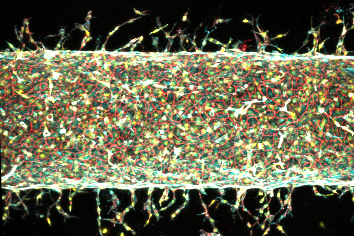
- Moderate Stretching (5% of gel width): When the gel was stretched back and forth by approximately 5 percent of its total width, a profusion of new vessels grew out from the main artery. This level of stimulation appeared to be optimal for maximizing vessel density.
- Increased Stretching (15% of gel width): Interestingly, when the stretching was increased to 15 percent, fewer new vessels sprouted. However, the vessels that did emerge tended to be longer, suggesting that the degree of mechanical force can influence not only the quantity but also the morphology and extent of vascular growth.
- Directional Guidance: Perhaps the most remarkable finding was the ability to direct the growth of new vessels. When the team changed the direction of the mechanical stretching, the newly sprouting capillaries responded in kind, following the applied pattern. This demonstrated a programmable control over vascular architecture, allowing researchers to essentially "draw" the path of new blood vessels using physical cues.
"The main takeaway is: Stretching the blood vessel back and forth seems to enhance the number of new capillaries that grow," Raman states. "We’re finding that moving is good, which is always the takeaway of everything we do in our lab. Mechanical forces play an important role in our bodies. That means that if you want to grow more or less vessels, or shorter or longer vessels, or vessels in certain directions, we now know how to do that." This statement encapsulates the paradigm shift offered by this research: moving beyond passive observation to active, directed control of vascular growth.
Unraveling the Mechanistic Link: The PIEZO1 Gene
While the "how" of mechanical stimulation was demonstrated, the "why" remained a crucial question. The researchers delved deeper to understand the underlying molecular mechanisms that translate physical force into biological response. Their attention turned to a specific gene: Piezo1.
The significance of PIEZO1 in mechanobiology was illuminated by the groundbreaking work of molecular biologist Ardem Patapoutian. In 2021, Patapoutian was awarded the Nobel Prize in Physiology or Medicine for his discovery of ion channels in cell membranes that open and close in response to mechanical pressure. These channels, named PIEZO1 and PIEZO2, act as cellular "gatekeepers," regulating the flow of ions (such as calcium) into and out of cells in response to physical deformation. Both types of channels are regulated by their respective genes. Patapoutian’s work fundamentally changed our understanding of how cells sense touch, pain, and proprioception (the sense of body position).
Raman’s interaction with Patapoutian after one of his talks proved pivotal. Upon seeing Raman’s experimental results demonstrating a clear link between mechanical stimulation and blood vessel growth, Patapoutian immediately proposed that the PIEZO1 channel could be the missing link. He hypothesized that the mechanical "exercise" applied to the central artery might be activating these PIEZO1 ion channels in the endothelial cells, thereby triggering the cascade of events that leads to new blood vessel formation.
To test this hypothesis, Raman’s team employed gene-editing techniques to suppress the PIEZO1 gene in the endothelial cells. The rationale was clear: if PIEZO1 was indeed the key mediator, then reducing its activity should diminish or abolish the mechanical stimulation-induced blood vessel growth. The results confirmed their suspicion. When the experiments were repeated with endothelial cells genetically modified to suppress the PIEZO1 gene, significantly fewer new blood vessels sprouted, even under the same regimen of mechanical exercise. This provided compelling evidence that blood vessels grow in response to mechanical stimulation specifically through the activation of PIEZO1 ion channels. This discovery not only explains the observed phenomenon but also opens new avenues for pharmacological interventions targeting these channels to modulate vascular growth.
Broader Implications and Future Horizons
The implications of this research extend far beyond the laboratory bench, promising to revolutionize several fields:
-
Regenerative Medicine and Artificial Organs: The most immediate and significant impact is on the development of functional engineered tissues and artificial organs. The ability to precisely pattern vascular networks could enable the creation of larger, thicker, and more complex tissues that can survive and integrate within the body. This could pave the way for laboratory-grown organs for transplantation, repairing damaged tissues after injury or disease (e.g., heart, liver, kidney, skin grafts for burn victims), and even creating vascularized tumor models for cancer research. Co-author Jessica Shah notes, "We are now investigating how precisely patterning blood vessel growth can help improve muscle function," highlighting the immediate practical applications already underway.
-
Drug Discovery and Disease Modeling: The "blood-vessel-on-a-chip" platform, now enhanced with programmable vascularization, offers a superior in vitro model for studying vascular diseases like atherosclerosis, diabetic retinopathy, and tumor angiogenesis. Researchers can create more physiologically relevant models to test new drugs, understand disease mechanisms, and observe drug efficacy and toxicity in a vascularized environment, potentially reducing reliance on animal testing.
-
Cancer Research: Angiogenesis, the formation of new blood vessels, is a critical process in tumor growth and metastasis. Tumors require a robust blood supply to grow beyond a few millimeters. The ability to control and understand angiogenesis through mechanical cues could offer new insights into tumor vascularization and potentially lead to novel anti-angiogenic therapies that target the physical microenvironment of the tumor, in addition to or instead of biochemical pathways.
-
Biofabrication and Biomanufacturing: This research introduces a new principle for biofabrication. Beyond traditional 3D printing or chemical gradients, mechanical forces can now be added to the toolkit for designing and constructing biological structures with unprecedented precision. This could lead to the development of new bioreactors and manufacturing processes for biological materials.
-
Fundamental Mechanobiology: The study deepens our understanding of fundamental biological processes, reaffirming the critical role of mechanotransduction in cellular function and tissue development. It adds another layer to the complex interplay between genes, biochemical signals, and physical forces that govern life.
Despite the significant progress, challenges remain. Scaling up these microfluidic systems to create vascular networks for full-sized human organs is a monumental task. Further research will be needed to understand the long-term stability and functionality of these mechanically induced vessels, their integration with host tissues, and their ability to withstand physiological pressures. However, the fundamental breakthrough of controlling vascular patterning through physical cues marks a pivotal moment.
This research, supported in part by the U.S. Department of War Army Research Office Early Career Program, PECASE Grant, and a Department of War DURIP Program Grant, represents a powerful testament to interdisciplinary collaboration, combining mechanical engineering, cell biology, and genetics. The team of MIT co-authors, including Sina Kheiri, Jessica Shah, Shashaank Venkatesh, and Roger Kamm, along with Peiyuan Chai and Ryan Flynn from Harvard University, has illuminated a new path forward in the quest to build and heal with biological precision. By demonstrating that "moving is good" for blood vessels, just as it is for muscles and nerves, this work promises to accelerate the realization of engineered tissues that can restore function and improve countless lives.