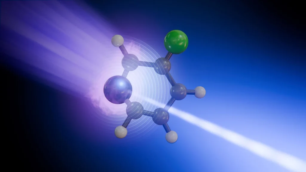
This groundbreaking research, conducted at the European XFEL, has provided an unprecedented atomic-level view of one of chemistry’s fastest and most fundamental processes: how molecules absorb light energy and subsequently redistribute it. For the first time, scientists have been able to discern the unique contributions of specific atoms within a molecule during this ultrafast transformation, demonstrating that not all atomic sites respond identically or relay the same information about the energy’s journey. The findings represent a significant leap forward in understanding light-driven chemical reactions, with profound implications for fields ranging from solar energy conversion to DNA photoprotection.
The Unseen Dance: Delving into Ultrafast Photochemistry
Photochemistry, the study of chemical reactions initiated by the absorption of light, underpins countless natural phenomena and technological applications. From photosynthesis in plants to the degradation of pollutants and the functioning of vision, light is a powerful driver of chemical change. However, the initial steps of these processes – the absorption of a photon, the excitation of electrons, and the subsequent structural rearrangements – occur on incredibly short timescales, often within femtoseconds (quadrillionths of a second) and picoseconds (trillionths of a second). Observing these fleeting dynamics in real-time, at the resolution of individual atoms, has long been a formidable challenge for scientists.
When a molecule absorbs light, its electrons jump to a higher energy state, making the molecule temporarily unstable. To return to a more stable state, this excess energy must be released or redistributed. This redistribution often involves a complex interplay between electronic motion and the movement of the atomic nuclei, leading to changes in the molecule’s shape and internal vibrations. A particularly crucial aspect of this process involves what scientists call "conical intersections." These are fleeting but critical crossing points on the molecule’s potential energy surface where different electronic states become degenerate, allowing for extremely rapid and efficient conversion of electronic energy into vibrational energy within the molecule’s atomic framework. Understanding these intersections is key to deciphering the efficiency and pathways of many light-triggered reactions.
Previous techniques have offered glimpses into these ultrafast events, but often lacked the atomic specificity needed to fully disentangle the complex dance of electrons and nuclei. The ability to precisely track energy flow and structural changes at specific atomic sites, as demonstrated in this new study, provides a richer, more nuanced picture than ever before. This level of detail is critical because the initial steps of a photochemical reaction dictate its entire subsequent course, influencing everything from reaction yield to product selectivity and energy efficiency.
Pioneering Technology: European XFEL and Time-Resolved Spectroscopy
The breakthrough was made possible by the cutting-edge capabilities of the European XFEL, located in Germany, which is the world’s largest X-ray laser. This facility generates extremely intense, ultrashort X-ray flashes that can penetrate matter and probe its atomic and electronic structure with unprecedented precision and speed. The X-ray pulses are so brief — lasting only femtoseconds — that they can effectively "freeze" atomic motion, allowing scientists to capture snapshots of molecular transformations as they unfold.
The specific technique employed in this research was time-resolved X-ray photoelectron spectroscopy (tr-XPS), conducted at the Small Quantum Systems (SQS) instrument of the European XFEL. tr-XPS is a powerful method that involves exciting a molecule with a pump pulse (in this case, an ultraviolet laser) and then probing it with a precisely timed X-ray pulse. The X-ray pulse ionizes the molecule by ejecting core-level electrons, and by measuring the kinetic energy of these emitted electrons, scientists can deduce the chemical environment around the specific atom from which they originated. Crucially, by varying the delay between the initial UV excitation pulse and the subsequent X-ray probe pulse, researchers can reconstruct a real-time "movie" of the molecular changes occurring over picoseconds.
The high brightness and ultrashort duration of the European XFEL’s X-ray pulses are indispensable for such experiments. The intense X-ray bursts provide enough signal from the sparsely distributed molecules in the gas phase, while their brevity ensures that even the fastest atomic and electronic motions can be resolved. This allows for the simultaneous observation of both electronic and structural changes with atomic resolution, a capability that has been a long-standing goal in physical chemistry.
Decoding Molecular Transformations: The Case of 3-Fluoropyridine
To demonstrate their novel approach, the researchers focused on 3-fluoropyridine, a relatively small, ring-shaped organic molecule containing nitrogen and fluorine atoms. This molecule was chosen for its manageability in experiments and its representative nature in exhibiting light-induced structural and electronic dynamics common to many more complex systems.
The experimental sequence began with a brief ultraviolet laser pulse delivering energy to the 3-fluoropyridine molecules. This absorbed energy excited the molecule’s electrons into a higher energy state. Immediately following this electronic excitation, the molecule quickly began to distort, bending out of its normally flat, planar structure. This structural change is a direct consequence of the altered electronic configuration, as the forces holding the atoms together shift.
As the molecule undergoes this rapid shape change, it passes through a critical region known as a conical intersection. As previously mentioned, these intersections are vital pathways in photochemistry, where the electronic and nuclear motions become strongly coupled. It is at these points that the molecule efficiently converts its excess electronic energy into vibrational energy within its atomic structure, effectively "cooling down" electronically while heating up vibrationally. Without these intersections, molecules would remain in excited electronic states for much longer, potentially leading to different, less efficient, or even damaging chemical reactions.
After traversing this intricate conical intersection, the molecule eventually returns to its ground electronic state, albeit now with significant vibrational energy. This excess vibrational energy then travels through the molecular structure, causing the atoms to oscillate and relax back towards their equilibrium positions over time. The ability to precisely follow this energy transfer and dissipation at an atomic level is where the new technique truly shines.
Atomic Storytellers: Fluorine and Nitrogen’s Unique Narratives
One of the most striking revelations of the study was the discovery that different atoms within the 3-fluoropyridine molecule provided distinct and complementary insights into the energy transformation process. This multi-site sensitivity is a game-changer for understanding complex molecular dynamics.
The fluorine atom, positioned on the periphery of the ring, served as a relatively clear and straightforward indicator. Its signal primarily reflected how the molecule’s vibrations relaxed and dissipated over time. As the absorbed electronic energy converted into atomic vibrations and then spread throughout the molecule, the fluorine atom’s environment changed in a way that directly correlated with the overall vibrational relaxation. This makes fluorine an excellent "reporter" atom for tracking the dissipation of vibrational energy, providing insights into the timescale and pathways of energy redistribution within the molecular scaffold.
In contrast, the nitrogen atom, which is an integral part of the molecule’s core ring structure and often plays a more direct role in electron density changes, told a much more intricate story. Because the nitrogen atom was more intimately involved in the initial electronic excitation caused by the UV light, its signal reflected a combination of both the redistribution of electrons and the molecule’s evolving structure. The X-ray photoelectron spectra from the nitrogen atom provided information about where the electron density was shifting and how the local bonding environment was changing as the molecule navigated its excited states and conical intersections. This dual sensitivity makes nitrogen a crucial sensor for understanding the fundamental electronic rearrangements that precede and accompany structural transformations.
Antonio Picón from the Instituto de Ciencia de Materiales de Madrid Consejo Superior de Investigaciones Científicas (ICMM-CSIC), a co-author of the study, emphasized this finding, stating, "We can now see that not every atomic site tells the same story in the signals we capture from our X-ray pulses. Some atoms report where the charge is going, while others reveal how the whole molecule vibrates." This insight highlights the power of site-specific probing, moving beyond a homogenized view of molecular dynamics to capture the individual contributions of each constituent atom.
Reconstructing the Picosecond Saga: Methodology and Data Interpretation
To achieve this unprecedented atomic-level view, the research team meticulously executed a series of experiments. First, the 3-fluoropyridine molecules were exposed to an ultrafast ultraviolet laser pulse, initiating the photochemical process. Following this, a carefully timed soft X-ray pulse from the European XFEL was used to probe the molecules. This X-ray pulse selectively ionized the molecules by removing deeply bound electrons from either the nitrogen or fluorine atoms, depending on the specific X-ray energy used.
The brilliance of the time-resolved approach lies in its iterative nature. The researchers repeated the measurement many times, systematically varying the delay between the initial UV laser pulse and the subsequent X-ray probe pulse. By recording the energies of the released electrons at each delay step, they were able to reconstruct how the chemical environment surrounding each specific atom — nitrogen or fluorine — changed over a timescale of just a couple of picoseconds (trillionths of seconds). This painstaking process allowed them to build a detailed, frame-by-frame "movie" of the molecular transformation.
However, the raw experimental data from tr-XPS is complex and requires sophisticated analysis. To fully interpret the experimental signals and connect them with the underlying electronic and structural changes within the molecule, the team relied heavily on advanced computer simulations and theoretical models. These computational tools were essential for translating the observed electron energy shifts into precise descriptions of electron density redistribution, bond length alterations, and overall molecular geometry changes. This synergistic approach, combining state-of-the-art experimental techniques with rigorous theoretical modeling, was critical for extracting meaningful chemical and physical insights from the ultrafast spectroscopic data.
Beyond the Molecule: Profound Implications for Science and Technology
While the experiment focused on a relatively simple molecule, the implications of this new methodology extend far beyond 3-fluoropyridine. The ability to track energy flow and atomic movements with such precision opens up a "new window on the microscopic mechanisms that govern photochemistry," as Daniel Rivas, former instrument scientist and now guest scientist at SQS and co-author of the study, noted. This fundamental understanding has profound potential across numerous scientific and technological domains:
-
DNA Repair and Damage: Understanding how DNA withstands exposure to ultraviolet light is crucial for comprehending mutagenesis and DNA repair mechanisms. Light absorption by DNA can lead to damage, but living organisms have evolved sophisticated repair pathways. By applying this technique, scientists could gain unprecedented insights into the ultrafast processes that lead to DNA damage or, conversely, to its photoprotection, potentially informing strategies for preventing skin cancer or developing novel therapeutic interventions.
-
Light-Harvesting Materials and Solar Energy: Improving the efficiency of solar cells and artificial photosynthesis hinges on a better understanding of how absorbed light energy is captured, transferred, and converted into useful work. This technique could be instrumental in designing more efficient organic photovoltaics, perovskite solar cells, or photocatalysts by revealing the atomic-level bottlenecks and pathways for energy transfer in these complex materials. Optimizing these initial energy conversion steps could lead to significantly more effective and affordable renewable energy technologies.
-
Photocatalysis and Industrial Chemistry: Many industrial chemical processes rely on catalysts activated by light. A detailed understanding of how light initiates reactions at the atomic scale could lead to the design of more selective and efficient photocatalysts for a wide range of applications, from synthesizing pharmaceuticals and fine chemicals to breaking down pollutants in water and air.
-
Drug Discovery and Photodynamic Therapy: Light-activated drugs are a growing area of pharmaceutical research. Understanding the precise molecular changes that occur when these drugs absorb light could lead to the development of more potent and targeted therapies, such as in photodynamic therapy for cancer, where light activates a drug to destroy cancer cells with minimal side effects.
-
Fundamental Chemical Physics: The study provides a deeper understanding of fundamental concepts such as electron-nuclear coupling, energy landscapes, and the role of quantum mechanics in chemical reactions. This detailed insight into the primary events of photochemistry will refine theoretical models and computational methods, pushing the boundaries of chemical physics.
A New Era for Photochemistry
The findings demonstrate how the unique capabilities of facilities like the European XFEL, with its ultrashort, high-brightness X-ray pulses, can separate and observe some of the fastest interconnected motions in matter. This multi-site sensitivity, combined with femtosecond resolution, represents a paradigm shift in how scientists can investigate photochemical processes.
The success with 3-fluoropyridine serves as a proof of concept, paving the way for applying this powerful approach to increasingly complex systems. Future research targets could include larger functional organic molecules, biomolecular building blocks like amino acids or nucleotides, and advanced materials designed for energy capture or transfer. By precisely mapping the energy flow and atomic rearrangements in these more intricate systems, scientists anticipate unlocking new design principles for materials with tailored photochemical properties.
As Daniel Rivas stated, "This is what European XFEL was built to enable: watching chemical change where it begins, at specific atomic sites and on its natural timescale." This research unequivocally fulfills that vision, launching a new era of direct observation in photochemistry and promising a cascade of discoveries that will impact fundamental science and technological innovation for decades to come. The ability to follow the distinct "stories" told by individual atoms as a molecule transforms under light provides an unparalleled narrative of chemical change, moving science closer to a complete understanding of the molecular world.