Materials known as relaxor ferroelectrics have been indispensable for decades, forming the bedrock of critical technologies spanning medical ultrasound imaging, high-fidelity microphones, and sophisticated sonar systems. Their remarkable performance, characterized by an unusual blend of high dielectric permittivity and electromechanical coupling, originates from the intricate and often enigmatic arrangement of atoms within their crystal lattice. For years, however, scientists have grappled with the immense challenge of directly measuring this internal atomic structure, forcing reliance on theoretical models that, while insightful, remained incomplete due to a lack of empirical validation at the atomic scale. This long-standing hurdle has now been decisively overcome.
In a landmark achievement poised to redefine the field of materials science, researchers from MIT, in collaboration with several esteemed institutions, have for the first time successfully mapped the three-dimensional atomic structure of a relaxor ferroelectric. Their groundbreaking findings, slated for publication in the prestigious journal Science, furnish an unprecedentedly clear foundation. This new understanding promises to significantly enhance and refine the models currently used to design and engineer future generations of computing systems, advanced energy devices, and highly sensitive sensors, heralding a new era of predictive materials design.
"Now that we have a better understanding of exactly what’s going on, we can better predict and engineer the properties we want materials to achieve," articulated James LeBeau, MIT’s Kyocera Professor of Materials Science and Engineering and a corresponding author of the study. He further emphasized the critical need for this breakthrough, stating, "The research community is still developing methods to engineer these materials, but in order to predict the properties those materials will have, you have to know if your model is right." The ability to validate theoretical constructs with direct atomic-scale observation marks a pivotal advancement, moving beyond mere hypothesis to confirmed structural understanding.
Unraveling the Enigma of Relaxor Ferroelectrics: A Historical Perspective
Relaxor ferroelectrics constitute a unique and highly important class of ‘smart’ materials, distinguished by their diffuse phase transitions and frequency-dependent dielectric response, particularly around their characteristic temperatures. Unlike conventional ferroelectrics which exhibit sharp, well-defined phase transitions, relaxors display a broad peak in their dielectric permittivity that shifts to higher temperatures with increasing measurement frequency. This distinctive behavior is attributed to the presence of nanoscale regions of local polarization, known as polar nanoregions (PNRs), which fluctuate in orientation and magnitude. These PNRs are typically embedded within a non-polar or weakly polar matrix, and their dynamic interactions are believed to underpin the materials’ exceptional electromechanical properties and energy storage capabilities.
The journey to understand relaxor ferroelectrics began in the mid-20th century, with the discovery of materials like lead magnesium niobate (PMN) in the 1950s and 60s. Early investigations quickly revealed their unusual dielectric properties, prompting intense scientific curiosity. For decades, researchers employed a battery of experimental techniques—including X-ray diffraction, neutron scattering, dielectric spectroscopy, and macroscopic electrical measurements—to probe their behavior. These methods provided invaluable insights into the macroscopic properties and average structural characteristics. For instance, neutron scattering helped confirm the existence of local structural distortions, hinting at the presence of PNRs. However, these techniques largely provided averaged information or projections, struggling to resolve the precise, three-dimensional atomic arrangements and the dynamic nature of PNRs at their fundamental length scales.
The inherent chemical disorder within relaxor ferroelectrics, often involving the substitution of different cations on specific crystallographic sites (e.g., B-site cations in perovskite structures like PMN-PT), contributes significantly to their complex behavior. This disorder leads to local variations in charge and strain, which in turn drive the formation and evolution of PNRs. Theoretical models, primarily based on statistical mechanics and first-principles calculations, have made considerable strides in describing these phenomena. Concepts such as the "random field model" and "superparaelectric model" emerged, attempting to explain the relaxor behavior by positing interactions between randomly distributed local electric fields and the fluctuating PNRs. Despite their sophistication, these models were ultimately constrained by the absence of direct, atomic-scale structural data for validation. The challenge was akin to trying to understand a complex, bustling city by only observing its traffic patterns and energy consumption, without ever seeing the buildings or the people within them.
A New Vision: Revealing Hidden Charge Patterns with Cutting-Edge Imaging
The breakthrough reported by the MIT-led team directly addresses this long-standing observational gap. In their meticulous study, the researchers deployed an advanced, state-of-the-art imaging method to scrutinize the distribution of electric charges throughout the relaxor ferroelectric material. What they ultimately uncovered fundamentally challenged and refined previous assumptions that had underpinned decades of theoretical modeling.
"We realized the chemical disorder we observed in our experiments was not fully considered previously," explained co-first authors Michael Xu, a PhD ’25 graduate, and Menglin Zhu, both postdocs at MIT. They elaborated on the iterative nature of their scientific process: "Working with our collaborators, we were able to merge the experimental observations with simulations to refine the models and better predict what we see in experiments." This synergistic approach, integrating high-resolution experimental data with computational refinement, proved crucial for advancing the understanding of these complex materials.
The interdisciplinary research team behind this monumental effort included a diverse group of experts. From MIT, Colin Gilgenbach and Bridget R. Denzer, both PhD students in materials science and engineering, contributed significantly. Further collaborations extended to Yubo Qi, an assistant professor at the University of Alabama at Birmingham; Jieun Kim, an assistant professor at the Korea Advanced Institute of Science and Technology; Jiahao Zhang, a former PhD student at the University of Pennsylvania; Lane W. Martin, a professor at Rice University; and Andrew M. Rappe, a professor at the University of Pennsylvania. This breadth of expertise underscores the collaborative spirit and multifaceted nature required to tackle such intricate scientific problems.
Probing Disordered Materials at the Atomic Scale: The Power of Multi-Slice Electron Ptychography
The core of this transformative research lies in the application of an advanced technique called multi-slice electron ptychography (MEP). For years, computer models had theorized that the remarkable energy storage and sensing capabilities of relaxor ferroelectrics stem from intricate interactions between positively and negatively charged atoms concentrated within these tiny, dynamic polar nanoregions (PNRs) when an electric field is applied. However, these nanoscale regions, typically just a few nanometers in size, had remained beyond the direct observational reach of conventional microscopy techniques.
To penetrate this veil of mystery, the researchers focused their efforts on a widely utilized relaxor ferroelectric material: a lead magnesium niobate-lead titanate (PMN-PT) alloy. This particular alloy is renowned for its exceptional piezoelectric properties, making it a staple in a myriad of high-performance applications, including medical transducers, industrial sensors, and advanced defense systems. The choice of PMN-PT as the target material was strategic, given its technological relevance and the long-standing questions surrounding its atomic-scale behavior.
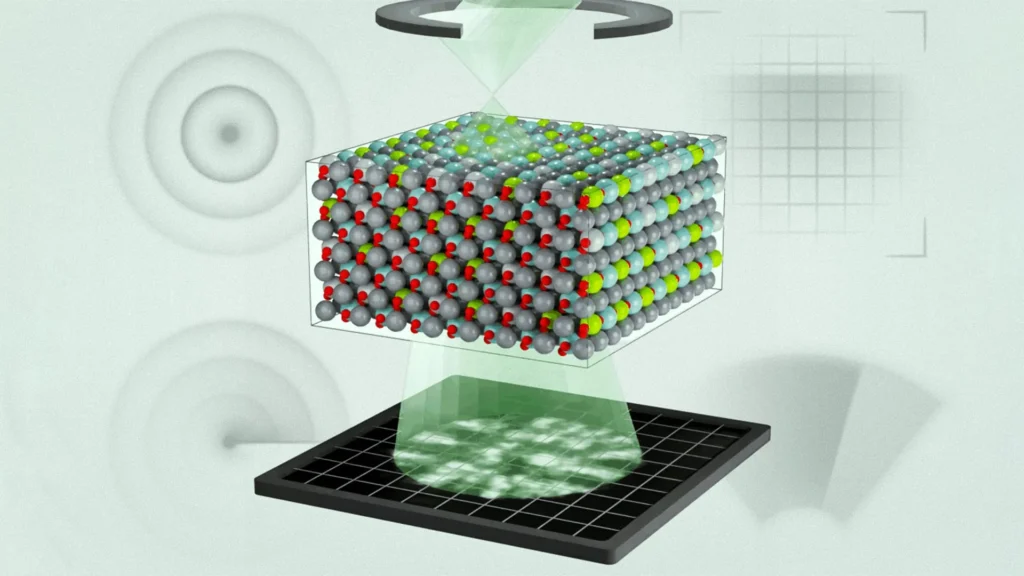
Multi-slice electron ptychography represents a significant leap forward in electron microscopy. It functions by systematically scanning a highly focused, nanoscale beam of high-energy electrons across the material sample. At each discrete position of the electron beam, the resulting diffraction pattern is meticulously recorded. "We do this in a sequential way, and at each position, we acquire a diffraction pattern," Menglin Zhu elaborated on the technical process. "That creates regions of overlap, and that overlap has enough information to use an algorithm to iteratively reconstruct three-dimensional information about the object and the electron wave function."
Unlike traditional electron microscopy techniques that primarily capture amplitude information, ptychography excels at recovering both the amplitude and phase of the electron wave that has interacted with the sample. This phase information is exceptionally sensitive to variations in the electrostatic potential within the material, making it ideal for mapping charge distributions and atomic positions with unprecedented precision. The iterative reconstruction algorithms, leveraging the redundancy of information from overlapping scan regions, allow for the generation of high-resolution, three-dimensional images of the atomic structure, even in complex, disordered materials where traditional imaging methods struggle. The ability to reconstruct the electron wave function provides a direct pathway to determining the projected atomic potential and, subsequently, the three-dimensional arrangement of atoms and their associated charge densities.
Key Discoveries and Model Refinement
Through the application of MEP, the team made several pivotal discoveries. They uncovered a sophisticated, layered hierarchy of chemical and polar structures within the PMN-PT alloy, extending from the precise positions of individual atoms all the way up to larger, mesoscopic features. This hierarchical organization provides a much more nuanced understanding than previously available, illustrating how atomic-level disorder propagates and influences larger-scale polar phenomena.
Crucially, the study revealed that the regions with distinct polarization were significantly smaller than earlier computer simulations had predicted. This finding directly challenges a core assumption in many existing theoretical models. By incorporating these experimentally validated observations—specifically, the refined understanding of chemical disorder and the actual size and distribution of polar regions—into their computational models, the researchers were able to dramatically improve how well simulations align with real-world material behavior. This iterative feedback loop between experiment and theory is a hallmark of robust scientific progress.
"Previously, these models basically had random regions of polarization, but they didn’t tell you how those regions correlate with each other," Xu explained, highlighting the qualitative leap in understanding. "Now we can tell you that information, and we can see how individual chemical species modulate polarization depending on the charge state of atoms." This insight into the atom-specific modulation of polarization is profound, as it directly connects the fundamental electronic structure and chemical composition to the macroscopic functional properties of the material. Understanding how the charge state of individual atoms influences the local polarization opens new avenues for precise control over material properties through atomic-scale engineering.
Broader Impact and Implications for Future Technologies
The findings from this study are not merely academic; they carry profound implications for the future of materials science and a wide array of technological applications. According to Menglin Zhu, this research vividly highlights the burgeoning power of electron ptychography as an indispensable tool for exploring the most complex and disordered materials, suggesting it could ignite entirely new lines of scientific inquiry.
"This study is the first time in the electron microscope that we’ve been able to directly connect the three-dimensional polar structure of relaxor ferroelectrics with molecular dynamics calculations," Michael Xu underscored, emphasizing the unprecedented integration of experimental observation with advanced computational modeling. He added, "It further proves you can get three-dimensional information out of the sample using this technique." This validation of MEP’s 3D capabilities for complex, disordered systems firmly establishes its utility for future materials characterization.
The team firmly believes that this innovative methodology could fundamentally transform how scientists approach the design of materials with precisely tailored electronic properties. Such advancements are critical for driving progress in numerous high-tech sectors:
- Memory Storage: Enhanced understanding of ferroelectric domains could lead to more efficient and durable ferroelectric random-access memory (FeRAM), a non-volatile memory technology with faster write speeds and lower power consumption than traditional flash memory. The ability to control polarization at the atomic level could enable higher density and more reliable memory devices.
- Sensing Systems: From advanced medical ultrasound probes offering higher resolution and deeper penetration to highly sensitive industrial sensors for non-destructive testing, the improved design of relaxor ferroelectrics will lead to superior performance. This also extends to sophisticated acoustic sensors, sonar systems for underwater navigation and detection, and advanced defense applications requiring robust and precise sensory capabilities.
- Energy Devices: Relaxor ferroelectrics are pivotal in high-energy-density capacitors, crucial for pulsed power applications and grid stabilization. A deeper understanding of their atomic structure will enable the development of materials with even higher energy storage capacities and improved efficiency, contributing to more sustainable and powerful energy solutions. Furthermore, their potential in solid-state cooling technologies, leveraging electrocaloric effects, could offer environmentally friendly alternatives to traditional refrigeration.
- Actuators: The superior electromechanical coupling of relaxor ferroelectrics makes them ideal for precision actuators used in microelectromechanical systems (MEMS), adaptive optics, and micro-robotics. Atomic-level design could lead to actuators with enhanced displacement, faster response times, and greater force generation.
Professor LeBeau encapsulated the broader scientific philosophy underpinning this work: "Materials science is incorporating more complexity into the material design process — whether that’s for metal alloys or semiconductors — as AI has improved and our computational tools have become more advanced." However, he issued a crucial caveat, reiterating the necessity of empirical validation: "But if our models aren’t accurate enough and we have no way to validate them, it’s garbage in garbage out. This technique helps us understand why the material behaves the way it does and validate our models." This sentiment highlights a critical challenge in the era of AI-driven materials discovery: the need for robust experimental verification to ensure that theoretical predictions translate into tangible, high-performance materials. The ability to directly observe and quantify atomic-scale structures provides the essential ground truth needed to build truly predictive and generative AI models for materials design.
The research received substantial financial backing, underscoring its strategic importance to national interests and scientific advancement. Support was provided in part by the U.S. Army Research Laboratory, which seeks advanced materials for defense applications; the U.S. Office of Naval Research, focusing on naval capabilities; the U.S. Department of War (likely referring to the Department of Defense, a common historical or journalistic shorthand for military research funding); and a prestigious National Science Graduate Fellowship, supporting promising young scientists. Furthermore, the work significantly leveraged the cutting-edge facilities at MIT.nano, a hub for nanoscale research and fabrication. This collaborative funding mosaic emphasizes the broad recognition of the research’s potential to yield transformative technological benefits and fundamental scientific insights.
In conclusion, the direct atomic-scale mapping of relaxor ferroelectrics represents a monumental leap in materials science. By finally peeling back the layers of atomic mystery that have shrouded these vital materials for decades, scientists are now equipped with the validated knowledge to engineer materials with unprecedented precision. This breakthrough not only redefines our understanding of relaxor ferroelectrics but also sets a new paradigm for the characterization and design of complex, disordered materials, paving the way for a future replete with advanced computing, energy, and sensing technologies.