A groundbreaking study led by researchers at the Massachusetts Institute of Technology (MIT) has unveiled a novel method for precisely controlling the growth and patterning of new blood vessels through simple mechanical stimulation. This discovery, detailed in the Proceedings of the National Academy of Sciences, represents a significant leap forward in the field of regenerative medicine, offering scientists an unprecedented ability to engineer artificial blood vessels and dictate their intricate growth patterns. By repeatedly jostling or stretching an artery, the team demonstrated that they could stimulate the sprouting of smaller capillaries, direct their growth trajectories, and even influence their number and length, thereby overcoming long-standing challenges in creating functional vascular networks within engineered tissues.
The research hinges on the elegant principle that mechanical forces, rather than solely biochemical cues, can serve as powerful architects of biological development. Ritu Raman, an associate professor of mechanical engineering at MIT and co-lead author of the study, emphasized the profound implications of these findings. "Healthy tissues depend on organized blood vessel networks, but state-of-the-art protocols don’t enable fabricating such networks within engineered tissues," Raman stated. "The ability to program blood vessel growth with physical cues may enable reproducible and scalable fabrication of engineered tissues that can be implanted in the body to restore function after debilitating disease or injury." This assertion underscores the potential for this technique to revolutionize the creation of implantable tissues and organs, where a robust and organized blood supply is paramount for survival and function.
The Critical Need for Vascularization in Regenerative Medicine
The quest to engineer functional human tissues and organs outside the body has been one of the most ambitious undertakings in modern medicine. From developing "organs-on-a-chip" for drug testing to fabricating replacement tissues for patients suffering from injury or disease, the promise of regenerative medicine is vast. However, a persistent and formidable hurdle has been the challenge of vascularization – the process of creating a dense network of blood vessels to supply these engineered tissues with oxygen and nutrients, and remove waste products. Without an efficient blood supply, engineered tissues, especially those exceeding a few hundred micrometers in thickness, rapidly become necrotic and non-functional.
Traditional methods for vascularizing engineered tissues have faced significant limitations. While advancements in 3D printing technology allow for the fabrication of larger arteries and veins, the intricate, microscopic networks of capillaries – the body’s most abundant and functionally critical blood vessels – remain exceedingly difficult to replicate with precision. Capillaries are responsible for the vital exchange of gases, nutrients, and waste at the cellular level, and their chaotic or insufficient growth can doom an otherwise promising tissue construct.
Scientists have also experimented with chemical cues, such as growth factors, to encourage blood vessel formation from individual cells cultured in Petri dishes. While these approaches can induce vessel growth, controlling the precise architecture, density, and directionality of these nascent networks has proven elusive. "You can try to pattern chemical cues, like growth factors, to direct where vessels grow, but you can’t do this very precisely," Raman explained, highlighting the inherent imprecision of purely biochemical guidance. This lack of control has severely hampered efforts to create clinically viable, complex tissues that mimic the sophisticated vascular organization found in native organs. The MIT team’s work directly addresses this critical gap by introducing a highly controllable physical patterning mechanism.
Building on Prior Success: The Power of Mechanical "Exercise"
The current breakthrough is not an isolated discovery but builds upon a foundation of previous research from Raman’s laboratory, which has consistently explored the profound impact of mechanical forces on biological systems. Her team previously developed protocols to grow artificial muscles and nerves, demonstrating that physical "exercise" or repeated mechanical stimulation could directly influence the growth and organization of these cell types. In those earlier works, the researchers designed a sophisticated chip containing a nutrient-infused gel. Embedded within this gel was a small magnet, and the gel’s surface was carpeted with live muscle or neuron cells. By manipulating an external magnet, the team could pull the embedded magnet and the cell-covered gel back and forth, observing how this mechanical activity guided cellular development and promoted functional organization.
This established success with muscle and nerve cells provided a compelling hypothesis: if mechanical forces could direct the growth of these tissues, could they also be harnessed to control the notoriously complex process of blood vessel formation? This question spurred the development of the "blood-vessel-on-a-chip" platform central to the new study.
The "Blood-Vessel-on-a-Chip": A New Paradigm for Vascular Control
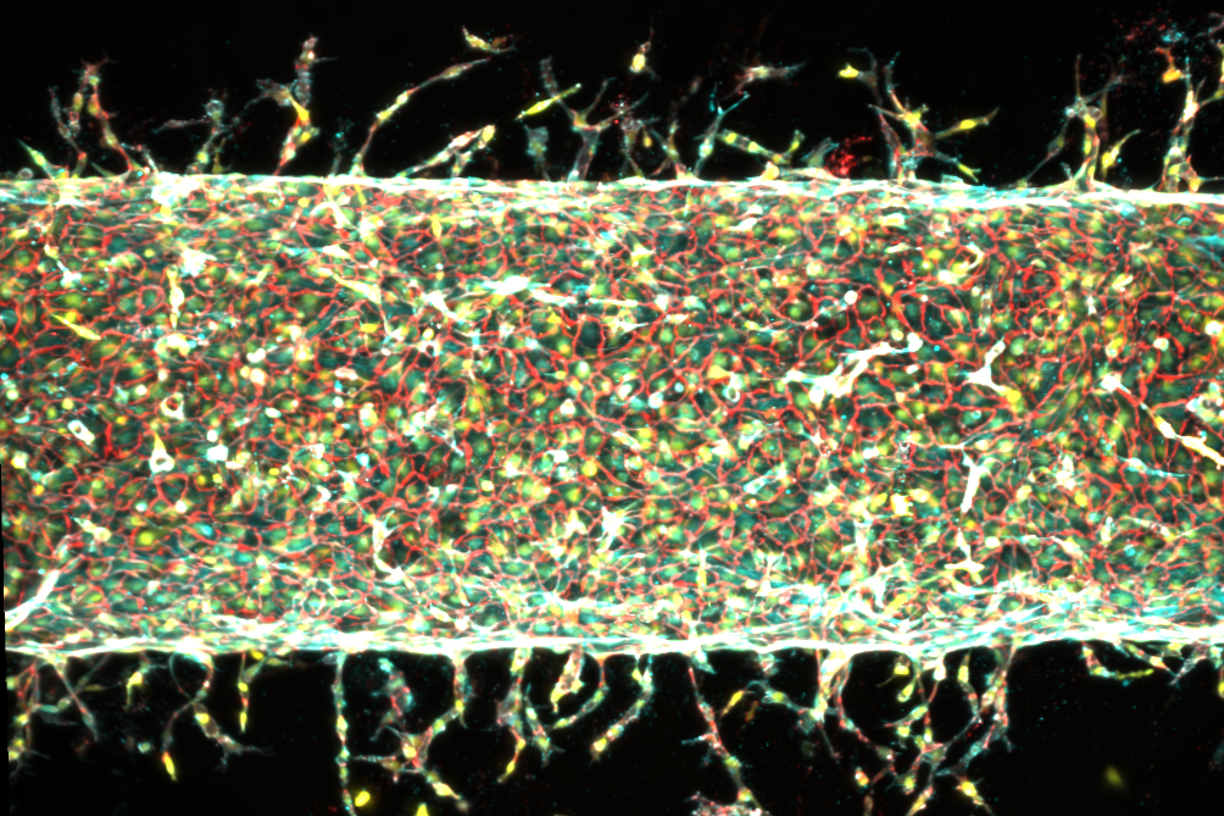
To test their hypothesis, the MIT team engineered a microfluidic device, aptly termed a "blood-vessel-on-a-chip," roughly the size of a postage stamp. This innovative platform was filled with a nutrient-rich gel, similar to their previous setups, and also contained a small magnet. A critical modification involved creating a thin, hollow channel lengthwise through the gel. This channel was then lined with live endothelial cells – the specialized cells that naturally form the inner lining of blood vessels throughout the body. Once these endothelial cells adhered to the channel’s shape, they began to mimic their natural behavior, sprouting new, capillary-like vessels into the surrounding gel.
The ingenious aspect of this setup lay in its precise control mechanism. The device was placed under a motorized stage equipped with small, suspended magnets. By meticulously moving these external magnets back and forth in different directions and with varying degrees of displacement, the researchers could apply controlled mechanical forces to the central, developing artery. They then carefully observed the response: whether and how new blood vessels sprouted from the main artery in response to these precisely applied mechanical stimuli.
The results were striking and unequivocally demonstrated the power of mechanical cues. In control experiments where the main artery was left undisturbed in the gel, some new vessels would sprout, but their location and directionality appeared random. However, when the artery was subjected to repeated mechanical jostling, a significantly greater number of vessels emerged. The team quantified these effects, finding that stretching the gel back and forth by approximately 5 percent of its total width resulted in a proliferation of new vessels branching out from the main artery. Interestingly, when the stretch was increased to 15 percent, fewer vessels sprouted overall, but those that did were noticeably longer. This demonstrates a nuanced control over both the density and morphology of the nascent vascular network.
Perhaps most remarkably, the researchers discovered that they could actively steer the direction of new vessel growth. By changing the direction of the mechanical stretching, the newly sprouting vessels dutifully followed suit, taking turns and conforming to the imposed pattern of mechanical stimulation. This unprecedented level of directional control represents a monumental step toward building organized, functional vascular trees.
Raman summarized these findings with a concise yet profound observation: "The main takeaway is: Stretching the blood vessel back and forth seems to enhance the number of new capillaries that grow. We’re finding that moving is good, which is always the takeaway of everything we do in our lab. Mechanical forces play an important role in our bodies. That means that if you want to grow more or less vessels, or shorter or longer vessels, or vessels in certain directions, we now know how to do that." This statement highlights the establishment of a programmable, physical toolkit for vascular engineering.
Unraveling the Mechanism: The Gatekeeping Role of Piezo1
Beyond simply observing the phenomenon, the research team delved deeper to understand the underlying biological mechanisms driving this mechanically induced vessel growth. Their investigation led them to a critical gene known as Piezo1.
The connection to Piezo1 was inspired by a talk Raman attended by molecular biologist Ardem Patapoutian. Patapoutian, a towering figure in the field of mechanobiology, was awarded the Nobel Prize in Physiology or Medicine in 2021 for his groundbreaking discovery of ion channels in cell membranes that open and close in response to mechanical pressure. These channels, named PIEZO1 and PIEZO2, act as cellular gatekeepers, regulating the flow of ions into and out of cells in response to physical deformation. Patapoutian’s work established that these channels are crucial for our sense of touch, proprioception (the sense of body position), and various other physiological processes where cells need to sense and respond to mechanical forces.
Following Patapoutian’s lecture, Raman shared her team’s experimental results, which clearly indicated a strong correlation between blood vessel growth and mechanical stimulation. Patapoutian, recognizing the parallels with his own research, astutely proposed that the PIEZO1 channel could be the missing link. He hypothesized that the mechanical "exercise" applied to the central artery in Raman’s experiments might be stimulating these ion channels in the artery’s endothelial cells to open, thereby triggering a cascade of intracellular events that ultimately led to the growth of new blood vessels.
To rigorously test this hypothesis, Raman’s team embarked on a gene-editing experiment. Their strategy was to "knock down" or suppress the activity of the PIEZO1 gene in the endothelial cells. If PIEZO1 was indeed the critical mediator, then a reduction in its activity should result in fewer blood vessels sprouting, even under mechanical stimulation. This would provide strong evidence that vessel growth in response to mechanical forces is specifically mediated through the activation of PIEZO1 ion channels.
The results were conclusive. When the experiments were repeated using endothelial cells that had been genetically engineered to suppress the PIEZO1 gene, the team observed a significant reduction in the number of new blood vessels that sprouted. This occurred despite the continued application of mechanical exercise to the central artery. This finding provided compelling evidence that PIEZO1 acts as a crucial mechanosensor, translating physical forces into biochemical signals that drive angiogenesis (the formation of new blood vessels).
Broader Impact and Future Implications
The discovery of a precise, programmable method for controlling blood vessel growth through mechanical stimulation, coupled with the identification of the underlying PIEZO1 mechanism, carries profound implications across several scientific and medical domains.
Firstly, this breakthrough is poised to accelerate the development of complex engineered tissues and artificial organs. One of the primary limitations in creating lab-grown organs like kidneys, livers, or hearts has been the inability to create a functional, integrated vascular system. The MIT team’s method offers a tangible path towards fabricating tissues with organized vascular networks that can be seamlessly integrated into the body, improving the success rates of transplants and regenerative therapies. For patients suffering from organ failure, severe injuries, or chronic diseases requiring tissue repair, this technology could eventually translate into more effective and durable treatment options.
Secondly, the enhanced control over vessel patterning could revolutionize "organ-on-a-chip" technologies. These microfluidic devices, designed to mimic the physiology of human organs, are increasingly used for drug discovery, toxicity testing, and disease modeling. By precisely controlling the vascular architecture within these chips, researchers can create more accurate and physiologically relevant models, leading to more efficient and reliable drug development, potentially reducing the need for animal testing.
Thirdly, the insights into the PIEZO1 pathway open new avenues for pharmacological intervention. Understanding how mechanical forces regulate angiogenesis through PIEZO1 could lead to the development of new drugs that modulate blood vessel growth. This could be beneficial in conditions where increased vascularization is desired, such as wound healing, tissue regeneration, or recovery from ischemic events (like heart attacks or strokes). Conversely, it could also offer targets for inhibiting unwanted blood vessel growth, as seen in diseases like cancer (where tumors rely on extensive vascular networks for growth) or age-related macular degeneration.
The study’s co-authors, including Sina Kheiri, Jessica Shah, Shashaank Venkatesh, and Roger Kamm from MIT, along with Peiyuan Chai and Ryan Flynn from Harvard University, contributed to the multidisciplinary success of this research. Jessica Shah, a co-author, articulated the immediate next steps: "We are now investigating how precisely patterning blood vessel growth can help improve muscle function." This indicates a clear translational focus, aiming to apply the controlled vascularization to enhance the performance and longevity of engineered muscle tissues, which could have significant implications for treating muscular dystrophies or severe muscle injuries.
While the research is currently at the laboratory stage, the reproducibility and scalability promised by a mechanical patterning approach suggest that it could transition more effectively into clinical applications than methods reliant solely on complex biochemical gradients. The elegance of using physical cues, which are robust and relatively easy to apply and control, offers a distinct advantage over the more fragile and diffusion-limited nature of chemical signaling in larger tissue constructs.
This work was supported, in part, by critical funding from the U.S. Department of War Army Research Office Early Career Program and PECASE Grant, as well as a Department of War DURIP Program Grant. Such support highlights the strategic importance of this research for both civilian medical advancements and military applications, such as treating combat injuries or developing advanced prosthetic interfaces.
In conclusion, the MIT team’s discovery marks a pivotal moment in tissue engineering. By demonstrating that mechanical forces can precisely guide the growth and patterning of blood vessels, they have unlocked a powerful new tool for regenerative medicine. This paradigm shift, from relying predominantly on chemical signals to harnessing the inherent mechanosensitivity of biological systems, promises to accelerate the creation of functional, vascularized tissues and organs, bringing us closer to a future where debilitating diseases and injuries can be effectively treated with engineered biological solutions. The ability to dictate the intricate dance of blood vessel formation, one mechanical jostle at a time, is set to redefine the landscape of medical innovation.